If you have been caring for horse residents for some time, you may be familiar with some or all of the anatomy covered in this resource. (An advanced resource is in our plans, so stay tuned!) However, if you are new to caring for horses, or just wish to refresh your memory, then this resource is for you. Caring for horse residents is challenging to say the least. A lot of time and resources go into providing quality care for horse residents. Having a general understanding of their basic anatomy can help you effectively communicate with your equine veterinarian. This is, of course, is an important part of ensuring residents are getting the appropriate care! It can be helpful for staff to know how to better describe locations of injuries or changes in the bodies of residents, and be able to readily supply the veterinarian with important details that can affect the course of action in emergency situations. Printing out a diagram and hanging it in the staff area can help keep the information handy. There are several diagrams in this resource covering basic body anatomy and basic hoof anatomy. After each diagram, there will be a glossary of terms used in the diagram to provide more clarity.
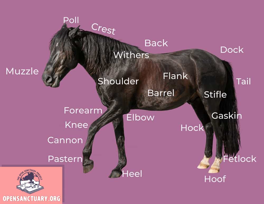
Let’s start out looking at a diagram showing basic horse anatomy. Knowing the vocabulary and the areas they refer to on a horse resident’s body will allow you to better understand your resident’s body, provide better care, and communicate more effectively with an equine veterinarian.
General Anatomy
Glossary Of Terms
Back
The back refers to the area that starts at the withers and ends at the last thoracic vertebrae. In Part 2 you will see the “loin” starts where the last thoracic vertebrae ends which will look like what we would consider part of a back.
Cannon
The cannon is the long, slender bone between the knee/hock and the fetlock.
Chin Groove
The chin groove is a dip behind the lower lip and chin.
Coronet
The coronet or coronary band is the ring around the where soft tissue meets the hoof. It is a very important area for the mobility of the horse.
Crest
The crest is the upper area of the neck.
Croup
The croup is the area that begins at the hip and stretches the length of the sacral vertebrae, ending at where the tail begins (the dock).
Dock
The dock is where the head meets the neck. It is located just behind the ears.
Elbow
The elbow is the joint of the front leg where the leg meets the body.
Ergot
The ergot is a callous, horn-like growth that may appear on one or all of the fetlock joints, along the backside. They are believed to be remnants of vestigial toes their ancestors had.
Fetlock
The fetlock is the joint connecting to the hoof. Sometimes called the “ankle” joint, it is more similar to the ball of the foot.
Forearm
The forearm is the area between the “knee” and the elbow.
Forehead
The forehead is the flat space between the eyes of a horse.
Gaskin
The Gaskin is an important muscle on the inside of a horse’s leg, just above the hock1: the tarsal joint or region in the hind limb of a digitigrade quadruped (such as the horse) corresponding to the human ankle but elevated and bending backward 2: a joint of a fowl's leg that corresponds to the hock of a quadruped area and below the stifle area.
Heel
The heel is the back side of the hoof.
Hock
The hock is a large, bending joint on the hind leg, below the gaskin muscle and above cannon bone.
Hoof
The hoof is the foot of the horse, consisting of a hard exterior and softer interior. Similar to a fingernail but much stronger.
Knee
The knee (carpus) is a large, bending joint of the front leg. It functions more like a wrist than a knee.
Muzzle
The muzzle consists of the nose, mouth, and chin of a horse.
Pastern
The pastern is the connecting area between the fetlock joint and the coronet (coronary band).
PollThe poll is the area where the head meets the neck, right behind the ears.
The poll is the area where the head meets the neck, right behind the ears.
Shoulder
The shoulder is the space under the withers, above the leg.
Stifle
The stifle is the knee of the hind leg, complete with a patella. This is where the leg meets the body.
Withers
The withers are just above the shoulder blades and are the highest point in a horse’s back. This is where horse height is measured. You can see them best when a horse has their head down.
Now that we have looked at some of the basic anatomical features of the horse, let’s focus in on the hoof!
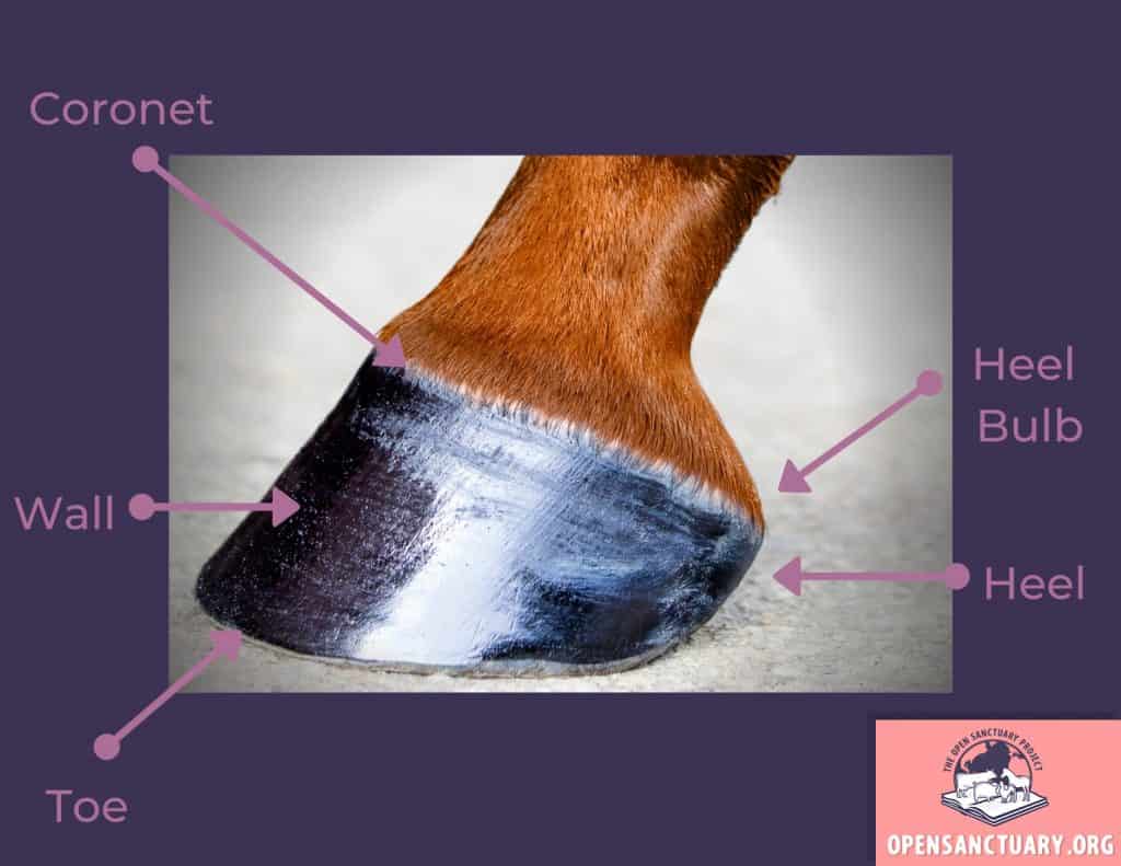
Basic Outer Hoof Anatomy
Glossary Of Terms
Coronet (Coronary Band)
The coronet or coronary band is the ring around the where soft tissue meets the hoof. It is a very important area for the mobility of the horse.
Heel
The heel is the back side of the hoof.
Heel Bulb
Heel bulbs are soft tissue at the back of the hoof that encloses the digital cushion (more on that in Part 2).
Toe
The toe is the front part of the hoof and the hoof wall.
Wall
The wall is the part of the hoof that bears weight and protects the inner hoof. It is made of keratin like human fingernails and grows from the coronet band.
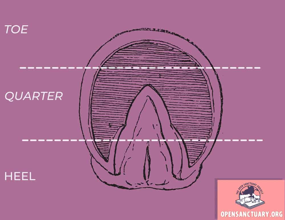
As you already know, there is a lot going on on the underside (solar) side of a horse’s hoof as well!
Basic Underside Hoof Anatomy A
Glossary Of Terms
Toe
The toe is the front part of the hoof.
Quarter
The quarter(s) are the sides/area between the toe and the heel.
Heel
The heel is the back side of the hoof.
But wait! There’s more to it than that.
Basic Underside Hoof Anatomy B
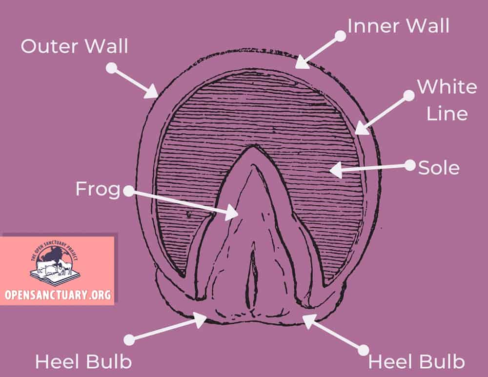
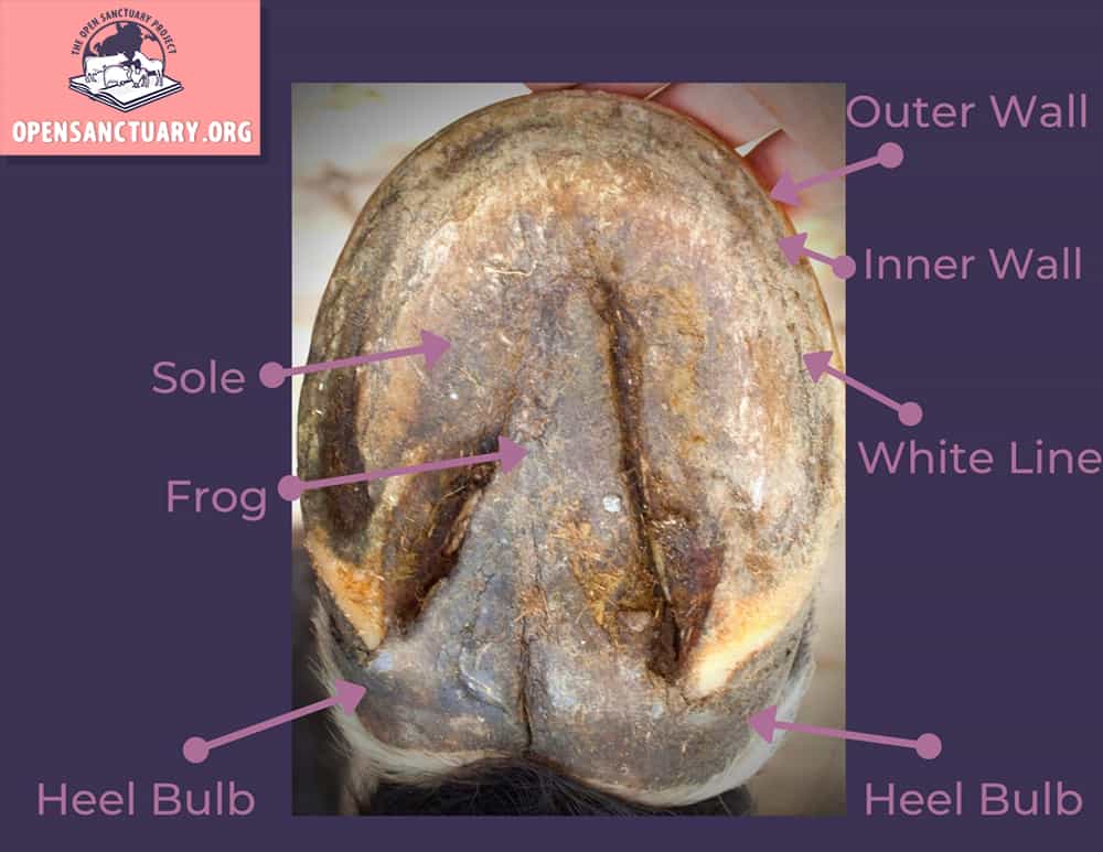
Easy enough, but what does this look like on an actual hoof?
Diagram Of Real Hoof
Glossary Of Terms
Frog
The frog is the thick, rubbery triangle or v- shaped structure in the mid/hind center of the hoof. It plays many important roles in the health of the hoof such as distributing impact and aiding in traction.
Heel Bulb
Heel bulbs are soft tissue at the back of the hoof that encloses the digital cushion (more on that in Part 2).
Inner Wall
The inner, or laminary, wall is the more pliable part of the wall, between the hard outer wall and the white line. Note the inner wall is actually whiter in color and the whiteline can appear more of a yellowish thin groove.
Outer Wall
The outer wall is harder and much stronger than the inner wall, protecting it and the hoof from damage.
Sole
The sole is the area within the white line that protects the inner structures but doesn’t include the frog or bars. (More on bars in Part 2.)
White Line
The white line is the area where the sole of the hoof connects to the wall. It forms a shallow groove on the bottom of the hoof and seals and protects the coffin bone. It is important to note it is actually more yellowish in color and the inner wall is whiter.
Well done! That’s a lot of information for basic horse anatomy. However, this knowledge can help provide better care for your horse residents and give staff a good foundation to build from. This resource is far from extensive and a “Part 2” will be in the works to help sanctuaries build upon their current knowledge or act as a refresher.
Non-Compassionate Source? If a source includes the (Non-Compassionate Source) tag, it means that we do not endorse that particular source’s views about animals, even if some of their insights are valuable from a care perspective. See a more detailed explanation here.
Article Tags
anatomy, basic horse anatomy, horse health
About Author
Amber Barnes
Amber is the Research Specialist of the Open Sanctuary Project.