If you have already cared for horses for some time, you may be familiar with some or all of the hoof anatomy covered in this resource. However, if you wish to expand upon your knowledge of basic hoof anatomy or to refresh your memory, then this resource is for you! If you are new to caring for horses, we recommend you check out the Basic Horse Anatomy resource too, which includes general hoof and body anatomy.
Caring for horse residents is challenging, to say the least, and caring for their hooves is imperative for good health! Having a general understanding of their hoof anatomy can help you effectively communicate with your equine veterinarian. It can be helpful to staff to know exactly how to describe hoof injuries or changes and be able to readily supply the veterinarian (or farrierSomeone who provides hoof trimming and care, especially for horses or cows) with important details that can affect the course of action in treating a malady.
Printing out a diagram and hanging it in the staff area can help keep the information handy. There are several diagrams in this resource covering basic hoof anatomy and more advanced hoof anatomy. After each diagram, there will be a glossary of terms used in the diagram to deepen your understanding. Stay tuned for the “Hoof Health And Care” resource! Healthy hooves are an important aspect of ensuring a happy horse!
General Anatomy Of The Hoof
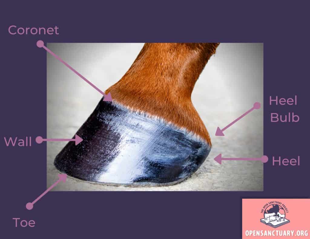
Let’s start by looking at the following diagram, which shows basic outer hoof anatomy. Knowing these words and the areas they refer to on a horse’s hooves will allow you to better understand your resident’s mobility, provide better care, and communicate more effectively with an equine veterinarian and farrier. Let’s start with a basic overview of the anatomy of the hoof and build upon that knowledge.
Side View Of Hoof
Glossary Of Terms
Coronet
The coronet or coronary band is the ring around the hoof, where soft tissue meets the hoof. It is a very important area for the mobility of the horse.
Heel
The heel is the back side of the hoof.
Heel Bulb
Heel bulbs are soft tissue at the back of the hoof that encloses the digital cushion.
Toe
The toe is the front part of the hoof and the hoof wall.
Wall
The wall is the part of the hoof that bears weight and protects the inner hoof, consisting of a hard exterior and softer interior. It is made of keratin, like human fingernails, but much stronger, and grows from the coronet.
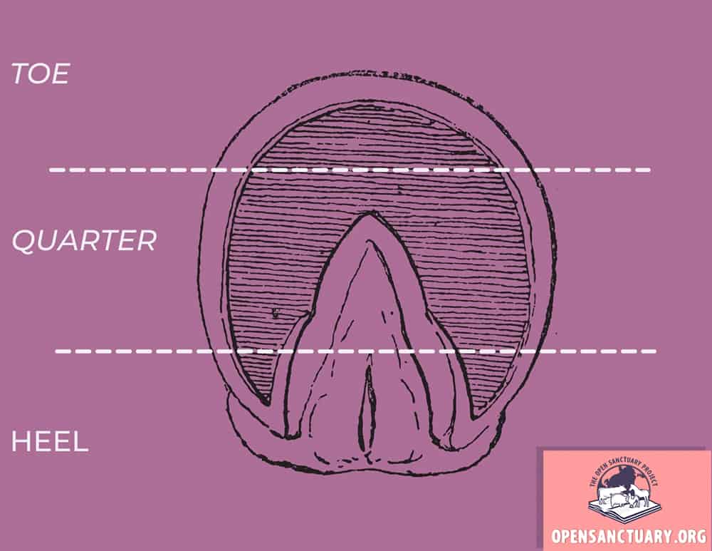
Main Sections Of Hoof
Glossary Of Terms
Toe
The toe is the front section of the hoof
Quarter
The quarter is the mid section of the hoof.
Heel
The heel is the back section of the hoof.
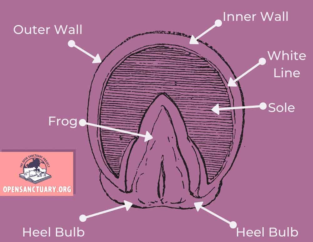
Bottom Of Hoof A
Glossary Of Terms
Frog
The frog is the thick, rubbery triangle or v-shaped structure in the mid/hind center of the hoof. It plays many important roles in the health of the hoof, such as distributing impact and aiding in traction.
Heel Bulb
Heel bulbs are soft tissue at the back of the hoof that encloses the digital cushion
Inner Wall
The inner, or laminary wall is the more pliable part of the wall, between the hard outer wall and the white line. Note the inner wall is actually whiter in color and the white line can appear more of a yellowish thin groove.
Outer Wall
The outer wall is harder and much stronger than the inner wall, protecting it and the inner structures of the hoof from damage.
Sole
The sole is the area within the white line that protects the inner structures, but doesn’t include the frog or bars. (Bars are a weight-bearing structure and part of the hoof walls that curve in and up. See diagram and glossary below.)
White Line
The white line is the area where the sole of the hoof connects to the wall. It forms a shallow groove on the bottom of the hoof and seals and protects the coffin bone. It is important to note it is actually more yellowish in color and the inner wall is whiter.
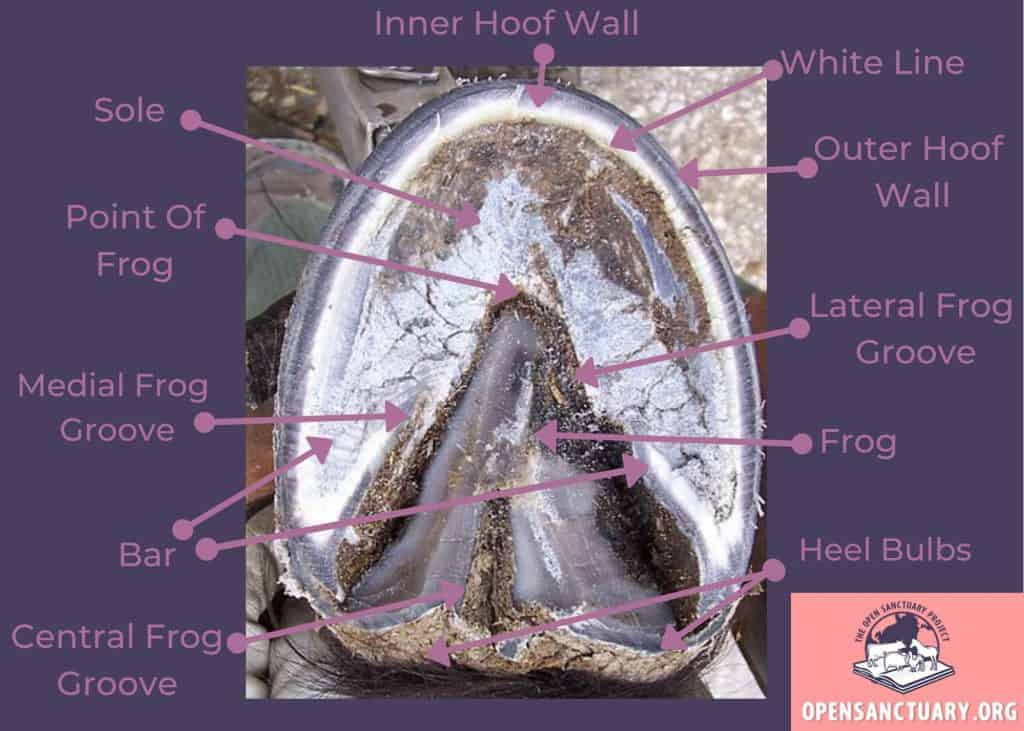
Bottom Of Hoof B
Glossary Of Terms
Bar
The bars are weight bearing structures and a continuation of the hoof wall that protrude from the heels towards the tip of the frog. They aid in shock absorption.
Central Frog Groove
The central frog groove, also referred to as the central sulcus, is a groove that runs a little ways up the center of the frog from the heel.
Lateral Frog Groove
Collateral grooves (or sulcus) are on either side of the frog. The lateral groove is towards the outer side of the hoof. Their depth runs the sole’s thickness. The grooves fill with dirt as the horse moves, which provides traction.
Medial Frog Groove
Collateral grooves (or sulcus) are on either side of the frog. The medial groove is towards the inner side of the hoof. Their depth runs the sole’s thickness. The grooves fill with dirt as the horse moves, which provides traction.
Point Of Frog
The point of the frog, also called the frog apex, is where the frog ends, narrowing into a tip.
Bottom Of Hoof C
Here they are all together. See what they look like on a real hoof. This hoof has been recently trimmed, making the walls and white line easier to distinguish.
Side View Of Internal Structures
Knowing even a few of the anatomical structures of the inner workings inside the hoof area can be especially beneficial as it helps you visualize healthy versus concerning hoof appearance and gaitA specific way of moving and the rhythmic patterns of hooves and legs. Gaits are natural (walking, trotting, galloping) or acquired meaning humans have had a hand in changing their gaits for "sport"..
Glossary Of Terms
Coffin Bone
The coffin (or pedal) bone determines how the hoof is shaped, with them shaped differently in the front and hind hooves. In both, it affects how the horse moves, allowing the horse to turn (hind hooves) and pull over the center of their hoof (front hooves).
Digital Cushion
The digital or plantar cushion sits above the frog in a hoof. It consists of springy tissues that dissipate shock during movement. It does this by expanding and contracting when the horse is moving.
Digital Flexor Tendon
This tendon attaches to the back of the coffin bone, allowing the horse to bend their pastern joints and their coffin joints.
Frog
The frog is the thick, rubbery triangle or v- shaped structure in the mid/hind center of the hoof. It plays many important roles in the health of the hoof such as distributing impact and aiding in traction.
Inner Wall
The inner, or laminary, wall is the more pliable part of the wall, between the hard outer wall and the white line. Note the inner wall is actually whiter in color and the white line can appear more of a yellowish thin groove.
Long Pastern Bone
The long pastern bone sits atop the short pastern bone, making up the pastern joint. When the horse moves, it absorbs the concussive impact.
Navicular Bone
The navicular bone rests behind the coffin bone and under the short pastern. The deep flexor tendon runs its way underneath the navicular bone and attaches to the coffin bone. It’s function prevents the coffin bone from over-extending.
Outer Wall
The outer wall encompasses the hoof and is harder and much stronger than the inner wall, protecting it and the hoof from damage.
Point Of Frog
The point of the frog, also called the frog apex, is where the frog ends, narrowing into a tip.
Short Pastern Bone
The short pastern bone sits under the long pastern bone, making up the pastern joint while acting as the top piece of the coffin joint. The ends of the bone are rounded, allowing the hoof to rotate on uneven ground.
Sole
The sole is the area within the white line that protects the inner structures but doesn’t include the frog or bars.
White Line
The white line is the area where the sole of the hoof connects to the wall. It forms a shallow groove on the bottom of the hoof and seals and protects the coffin bone. It is important to note it is actually more yellowish in color and the inner wall is whiter.
You made it! You can now use this knowledge can help provide better care for your horse residents and give staff a good foundation to build from. This resource is far from extensive and a hoof health and care resource will be in the works to help sanctuaries build upon their current knowledge or act as a refresher.
Non-Compassionate Source? If a source includes the (Non-Compassionate Source) tag, it means that we do not endorse that particular source’s views about animals, even if some of their insights are valuable from a care perspective. See a more detailed explanation here.
Article Tags
hoof anatomy, hoof health, hoof structure
About Author
Amber Barnes
Amber is the Research Specialist of the Open Sanctuary Project.