


Updated October 13, 2020
When it comes to horses, you’ll want to ensure that you catch and treat any health challenges as early as possible. To do this, you’ll need to spend a lot of time observing and getting to know your residents to better catch less obvious signs of concern. By conducting regular full body health evaluations, you’ll be able to know what healthy looks and feels (and smells!) like, and when you should be concerned.
To catch and respond to health issues as early as possible, you’ll need to spend a lot of time observing and getting to know your residents to better catch less obvious signs of concern. By conducting regular full-body health evaluations, you’ll be able to know what healthy looks and feels (and smells!) like, and when you should be concerned. Check out our guide to turkeyUnless explicitly mentioned, we are referring to domesticated turkey breeds, not wild turkeys, who may have unique needs not covered by this resource. health checks to familiarize yourself with the signs that something may be amiss with a horse resident. For more information on health challenges commonly affecting older horses, check out our resource here.
Issues By Body System
Circulatory: Equine Infectious Anemia (“Swamp Fever”)
Gastrointestinal: Anthrax, Colic, Constipation, Diarrhea, Enteroliths, Gastric Ulcers, Internal Parasites – Large Strongyles, Small Strongyles, Lungworms, Roundworms, Pinworms, and Tapeworms
Metabolic: Equine Cushing’s Disease, Equine Metabolic Syndrome, Insulin Resistance
Musculoskeletal System: Arthritis, Club Foot, Exertional Rhabdomyolysis, Hoof Abscesses, Hoof Avulsion, Hoof Cracks, Hoof Overgrowth, Immune-Mediated Myositis, Laminitis, Pigeon Fever, Thrush, White Line Disease (‘Seedy Toe’)
Neurological: Equine Encephalomyelitis, Equine Protozoal Myeloencephalitis, Tetanus (‘LockJaw’), West Nile Virus, Rabies
Reproductive System: Cryptorchidism
Respiratory: Anthrax, Equine Herpes Virus, Equine Influenza, Lungworms, Respiratory Airway Disease “Heaves”, Strangles
Skin And Hair: Abscesses, Anthrax, Brucellosis, Dermatophilosis (‘Rain Scald’), External Parasites ( Lice, Mites, and Ticks), Insect Hypersensitivity (‘Sweet Itch’), Pastern And Heel Dermatitis (‘Mud Fever’), Pigeon Fever, Ringworm, Sarcoids
Urinary System: Cystitis and Pyelonephritis
Vision: Cataracts, Conjunctivitis
Weight and Diet: Hyperlipaemia, Laminitis
Abscesses
An abscess occurs when white blood cells accumulate in response to a foreign body or infection, forming pus, and the body walls off the area with other cells. Abscesses can occur anywhere in a horse’s body, including the internal organs. More often, abscesses form in the hoof or under the skin where the pressure often builds up, rupturing the abscess. This rupturing can expel surprising amounts of pus, depending on the size of the abscess. After an abscess ruptures, the wound may continue to leak pus and become a chronic sore. Abscesses of the hoof are commonly caused by the puncturing of the hoof by something sharp that stays lodged within the hoof. In this case, the infection can grow into the deep tissues causing lameness. An example of abscesses of the internal organs is Strangles, an infection caused by Streptococcus equi that causes abscesses in the lymph nodes below the ear and under the throat, as well as in the internal organs.
If you suspect an abscess, call a veterinarian to diagnose it. They may want to do xrays They may apply a poultice to draw out the abscess to the surface or recommend you soak there hoof once or twice a day in a bucket of They may also lance the abscess and irrigate the wound (any abscess on the face or neck should be treated by a veterinarian to minimize risk of major bleeding). Ask them to take a sample of the pus to discover the type of bacteria causing the infection and dictate the type of antibiotics, if any, are required. This will also help you know if you need to isolate the horse to prevent the herd from becoming infected.
In the event that you do not have access to a veterinarian, lancing an abscess is a relatively simple process, though this technique should be taught by an expert or veterinarian prior to attempting! You can start by applying a poultice – heated, moist cloth to the site of the abscess. Then trim the hair around the abscess, disinfect the surface with an antiseptic, and make a low, small, vertical incision with a sharp and sterilized knife. Using sterile gloves, carefully squeeze out the excess pus and flush the wound with disinfectant. Discard or sterilize anything that comes into contact with the pus and monitor the wound for up to a month. You may want to isolate the horse depending on the abscess size or location for a time period. Certain diseases will require additional care and treatment of the abscessed horse. (Back to the top)
SOURCES:
The Working Equid Veterinary Manual | The Brooke
Anthrax
Anthrax is caused by Bacillus anthracis spores, which can lie dormant in soil across the world for many years. The bacteria can activate and contaminate soil and grass in certain weather conditions, especially wet and cool weather followed by hot and dry weather. Animals that graze are susceptible to the disease after eating contaminated grass. It can also be contracted through direct contact of anthrax with a lesion, nasal discharge, inhalation, and biting insects. Symptoms include high fever, chills, colic, depression, incoordination, staggering, trembling, convulsions, swollen neck, belly, or throat, increased respiratory rate, bleeding from the rectum, and unfortunately, typically death within just a few days. If you suspect a horse is suffering from anthrax, you must contact your veterinarian immediately. Anthrax can quickly spread to other animals from the infected horse, including humans. Confirmations of anthrax must be reported to government officials. There is also a vaccine available for anthrax. (Back to the top)
SOURCE:
Diseases That Affect Horses, Ponies, Mules, And Donkeys | National Center For Foreign Animal And Zoonotic DiseaseAny disease or illness that can be spread between nonhuman animals and humans. Defense (Non-Compassionate Source)
Anthrax Facts | American Veterinary Medical Association
Arthritis
Like most animals, horses can become prone to arthritis as they get older. Arthritis can also be caused by injury, infection, malnutrition, and a lack of space to move freely. Symptoms include pain and/or swelling in joints, reluctance to move, stiffness, laying down more often, weight loss from poor appetite, shabby coat (poor nutrition), strange gait, and signs of lameness. Treatment for arthritis differs depending on the root cause, so if you believe that a horse is suffering from arthritis, it’s important to consult with your veterinarian. Make extra sure that their environment is as arthritis-friendly as can be, minimizing steep grades or long walks to food or water if you can! (Back to the top)
SOURCES:
Arthritis In Horses | VCA Hospitals
Brucellosis
Brucellosis in equines generally presents as one or two conditions, namely poll evil and fistulous withers.
PollThe poll is the area where the head meets the neck, right behind the ears. evil is a disease where the area is injured or infected and becomes inflamed and swells, and eventually the infection leads to devitalized tissue.
Fistulous withers is a condition found in horses and donkeys where the supraspinous bursa (located near the withers) becomes inflamed. This condition can be caused by traumatic injuries or infectious agents. When an infectious agent is involved it is most often caused by Brucella abortus. Common signs of fistulous withers are swelling, pain, and heat, but can also include fever, lethargy and stiffness. Over time, the bursa may rupture and have an infectious looking discharge. This may heal over, leaving a scab, or it may continue to drain for many days. If left untreated, the problem can heal and then show up again some time later. In almost all cases, the area surrounding the bursa becomes thickened with scar tissue and inflammation.
If the bursa is un-ruptured and not draining, treatment with antibiotics and anti-inflammatory agents is recommended. When the bursa is fistulated or draining, most treatments include removing any involved tissues and flushing the area with dilute betadine or other solutions. Antibiotics are also often given to help prevent additional infections. Affected horses typically do not give this disease to other animals or humans, but it can be spread by cows, goats, wild pigs, sheep, deer, and related animals to horses, donkeys, and humans. Keep horses away from infected cows, sheep or goats and keep infected horses with openly draining fistulous withers separate from other residents. (Back to the top)
SOURCES:
Equine Brucellosis: Review On Epidemiology, Pathogenesis, Clinical Signs, Prevention And Control | Journal Of Experimental Biology And Agricultural Sciences (Non-Compassionate Source)
Diseases With Horse To Human Transmission | UC Davis Center For Equine Care (Non-Compassionate Source)
Cataracts
A cataract is an increased density or opacity of an eye’s lens, reducing the transmission of light to the retina. Some cataracts are congenital, while others are acquired. Acquired causes of cataracts can be caused by trauma or ocular disease, UV light, toxin ingestion, and ionizing radiation. Aging horses may also develop senile cataracts. Cataracts may appear as an initial grayness, or bubbles or cracks in the lens. These progress to white or grey opacity, preventing light from being reflected from the retina. This obscures the structures behind the lens. It is important to treat the underlying conditions and associated symptoms. Unfortunately, cataracts are irreversible once formed. Only specialized surgery can potentially fix the issue. In cases where vision is limited, it is important to ensure the affected horse is in a safe place without environmental dangers. (Back to the top)
SOURCES:
The Working Equid Veterinary Manual | The Brooke
Conjunctivitis
Conjunctivitis is a symptom or response of the eye to irritation, illness, or injury. It is often referred to as Pink Eye. The conjunctiva includes the inner eyelid and surrounding membrane. There are many cause of conjunctivitis, ranging from viral disease to fungal and protozoal infections to allergies, or even cancer. Often conjunctivitis is caused by an irritant in the eye, such as dust, flies, or a foreign body. Trauma to the eye can also cause conjunctivitis. Conjunctivitis presents as a reddened, inflamed membrane, and is sometimes accompanied by swelling or watery/mucousy discharge.
Washing the eye to remove dirt or a foreign object is a good first step. It’s important to identify the cause as it will alter the course of treatment. Call a veterinarian to discuss the symptoms and proper course of treatment. (Back to the top)
SOURCES:
The Working Equid Veterinary Manual: Chapter 9 | The Brooke
Constipation
Constipation is an uncomfortable and, sometimes serious, condition (impaction colic). There are a number of factors that can lead to constipation including poor dental health, ingestion of sand or foreign objects, certain medications, intestinal damage caused by parasites, lack of water, gastrointestinal disease, or poor diet. You may notice a resident straining when defecating or find hard, dry stools or mucous covered stools. You may notice an absence of manure all together.
To ensure a healthy intestinal tract, feed residents plenty of forage and keep grain to a minimum and provide constant access to water. If you have a resident who is prone to constipation, a veterinarian or equine nutritionist might recommend adding more fibrous components to their diet like beet pulp or bran. Because some causes are not diet related, it is important to consult your veterinarian about prevention or treatment options and whether the resident requires special veterinary care. They may suggest a horse-safe laxative such as milk of magnesia or psyllium with mineral oil for mild constipation or recommend dioctyl sodium sulfosuccinate in a powdered form, for hard stools. Constipation can indicate impaction colic which is a serious condition and requires immediate veterinary care. This may require intravenous fluids to be administered and a stomach tube for the ingestion of electrolytes or even surgical intervention. (Back to the top)
SOURCES:
The Working Equid Veterinary Manual Chapter 11 | The Brooke
Cystitis And Pyelonephritis
Cystitis (urinary tract infections involving the bladder) are common in equines. They are often associated with disorders that disrupt urinary flow. When a UTI has spread to the kidneys it is called pyelonephritis. Common organisms involved in these issues are E. coli, Enterococcus app., and Streptococcus app. Physical conditions that can lead to cystitis (and pyelonephritis) include calculi, urinary retention, neurological diseases, tumors, You may notice a horse frequently attempting to urinate, urinating frequently in small amounts, or notice blood in their urine. Other symptoms include dribbling or scalding of the perineum and legs. You may notice them losing coordination, appearing depressed, and showing little interest in food. Cytitis/pyelonephritis is often accompanied by a fever.
Horses that have had multiple catheters placed, have had obstructions of the bladder/urinary tract, or are in late stages of pregnancy may be predisposed to UTIs. It is important to keep your horse well-hydrated and call a veterinarian to diagnose the issue and prescribe the appropriate treatment. (Back to the top)
SOURCES:
The Working Equid Veterinary Manual Chapter 13 | The Brooke
Club Foot
Club foot occurs when the coffin bone stays partially flexed due to the shortening of the deep flexor tendon and can’t be fully extended. Club foot, an abnormally shaped upright foot, can be caused by congenital or acquired deformities of the deep digital flexor tendon. It can also be caused by chronic limb pain while a hoof is treated, the disuse of the limb may cause the tendon to contract, pulling the pedal bone into a more upright position. It is important to identify the cause, removing the source of pain when possible. A veterinarian may recommend the use of NSAIDs but you must consult with them first to determine the cause and appropriate treatment. If the club foot is chronic, have the hoof trimmed regularly by a professional to keep the foot balanced. Although it may seem like a good idea to trim them into a “normal” shape, do not do this as it can cause lameness. Work closely with a veterinarian and a farrier to treat this condition. Be aware that toe abscesses are common and their living area should be free from sharp stones. (Back to the top)
SOURCES:
The Working Equid Veterinary Manual Chapter 11 | The Brooke
Colic
Colic isn’t so much a particular disease but the symptom of abdominal pain. Colic is a serious issue (even fatal) among horses and quick action is vital if you suspect colic. Always call a veterinarian if you suspect colic. There are many causes of colic. Various types of colic may present differently, and they are often assessed on the basis of the horse’s history, presence of pain, heart-rate, respiratory-rate, gut sounds, the presence/absence, frequency, and moisture level of stools, and other exam findings. While some cases of colic are mild and resolve, other cases can be fatal and all cases should be treated seriously.
When colic strikes, the intestines and/or the stomach may be inflamed, the intestinal walls may be distended, the motility of the intestines may be altered, and/or the intestines may be experiencing a loss of blood supply due to torsion. Colic can be a broken down into broad categories:
- Spasmodic colic
- Spasmodic colic can be caused by sudden changes in diet or nervousness, among other things.
- Impaction colic
- Large amounts of food that has larger pieces of unchewed forage can cause an impaction as can a lack of water, ingestion of foreign objects or sand.
- Displacement/Torsion colic
- This can happen if a loop of intestines becomes trapped or twisted. If twisted, this can result in a loss of blood supply.
- Excessive fermentation colic
- This can happen from eating too much grain that ferments in the stomach faster than it can eliminate it.
Symptoms:
- Depression
- Inappetence (not interested in eating)
- Pawing
- Looking at the flank
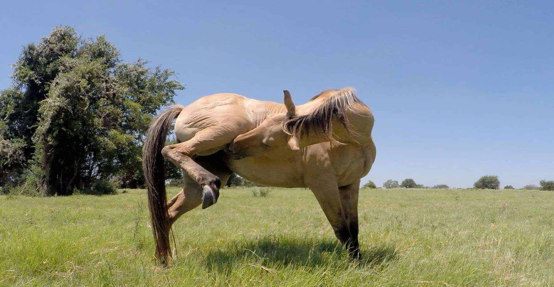
- Lying down more than usual or at a different time from normal (Figure 1)
- Lying down, getting up, circling, laying down again repeatedly
- Curling/lifting the upper lip
- Kicking up at the abdomen with hind legs
- Rolling up onto back
- Standing stretched out
- Sitting with hind legs on the ground
- Groaning
- Sweating
- Increased heart rate (normal range is 25–45 beats per minute)
- Visible abdominal distention (appearance of being bloated)
- Less than normal to no manure production
- Diarrhea
- Foals may roll up on their backs or grind their teeth and salivate excessively
If you suspect a horse is colicking, call your veterinarian immediately.
While you wait for the veterinarian to arrive, continue monitoring the unwell horse for further signs of discomfort. The veterinarian will perform an examination in order to accurately diagnose the issue. Depending on the findings of the examination, the veterinarian may decide to introduce fluids into the horse’s stomach via a tube inserted into a nostril. They may also put the horse onto a ‘drip’ (fluid introduction via the large vein in the neck) and prescribe pain-killers and antibiotics when appropriate. Sometimes hospitalization and/or surgery may be required. Unfortunately, there are scenarios where euthanasia may be the most compassionate option in grievous cases.
Possible Causes And Management:
- Feed – sudden changes to diet, poor quality food, too much grass, feeding cereals:
- Make any dietary changes gradually over at least a week, ideally 2-4 weeks. Feed good quality forage and horse specific proprietary foods. Avoid moldy food
- Always soak sugar beet to the manufacturer’s recommendations
- Ensure regular feeding: small amounts and often, especially if the animal is supposed to be eating extra calories
- Avoid access to too much rich spring grass, which can lead to problems with laminitis and colic
- Avoid access to grain and other rich food. Rich food, particularly those that are high in starch and sugar, can cause laminitis and colic
- Inadequate/dirty water supply:
- Check troughs at least daily: Self-fill auto-waterers can become blocked, water supply can fail
- Clean any contaminated water containers, as horses may not drink dirty water
- Check that water is not frozen or too cold. Some horses may not drink very cold water
- Consider offering several sources of water
- Eating non-food items such as plastic bags, rope or bedding:
- Ensure horses cannot access such material
- Watch out for horses eating their bedding, eg when box-rested under veterinary instruction
- Ingestion of poisonous plants:
- Know about the poisonous plants and trees that could be present at your sanctuary and prevent your residents from accessing them
- Check pasture and boundary fences and hedgerows frequently and remove them or fence off the problem area
- Fence off trees during fruiting to prevent overeating
- Sandy soil: Avoid grazing on sandy soil pasture if possible
- Dental disease – failure to chew food adequately resulting in a blockage of the gut:
- Ensure horse’s teeth are checked at least annually by a qualified equine dental technician or veterinarian following a dental care program
- Dental disease is more common in older horses. Suspect teeth problems if horses are ‘quidding’ (dropping partially-chewed food) or drooling saliva
- Parasites – migrating worm larvae or large numbers of worms causing an obstruction:
- Ensure regular fecal worm egg counts are carried out to determine if horses require treating for worms. Consult your veterinarian for advice
- Pick up manure from the paddock a minimum of twice per week
- Stomach ulcers:
- Reduce stress and ensure you ‘trickle feed’ the horse
- Pancreatitis:
- Tumors:
- If a tumor is the cause, surgical intervention may be necessary.
- Torsion:
- If the intestines are twisted, surgical intervention may be necessary.
SOURCES:
Colic | Stillwater Equine Veterinary Clinic
The Working Equid Veterinary Manual Chapter 11 | The Brooke
Cryptorchidism
Cryptorchidism is a condition where one or both testes fails to descend from the abdomen into the scrotal sac during the final weeks of gestation. Both testicles should be located within the scrotum when the male foal is born. A horse with cryptorchidism may remain fertile and this could lead to pregnancies in the resident population. Generally a horse with bilateral cryptorchidism will be sterile while a horse with unilateral cryptorchidism is usually still fertile. Horses with cryptorchidism (sometimes referred to as rigs) often display stallion-like behavior. If your veterinarian suspects cryptorchidism, there are several tests that can be done to confirm this diagnosis. Depending on the results of these tests, your veterinarian may recommend surgery. The type will depend on the area the undescended testes sits. (Back to the top)
SOURCES:
Cryptorchid Fact Sheet | The Dick Vet Equine Centre
Cryptorchidism (Undescended Testicles) In Horses | American College Of Veterinary Surgeons
Cushing’s Disease (Pituitary Pars Intermedia Dysfunction)
Cushing’s disease is a hormonal disorder that arises when the nerve supply to the pituitary gland becomes damaged. This ultimately causes increased quantities of hormones to be released from the gland. The disease is more correctly termed ‘Pituitary Pars Intermedia Dysfunction (PPID).’ the severity does seem to increase with age. There is also a possibility that obesity predisposes to PPID, but this has not been confirmed. Cushing’s is more likely to hit horses over 10 years old, though younger horse can certainly develop it. Nineteen years is the average age for horses to be diagnosed with PPID. While all equines can develop donkeys are particularly susceptible.
It is common to see signs of PPID in horses in the form of long, hairy coats, laminitis, increased sweating, and excessive urinating and drinking. Because PPID also affects body fat and muscle, a horse may present with a “potbelly” and lethargy. Diagnosing PPID can involve a number of tests and getting a good history and recording clinical signs.
While there is currently no cure for PPID, there are long-term treatment options available. Daily medication and bi-annual blood test are required for maintenance. There are several treatment options a vet may consider (dopamine agonists, serotonin antagonists and cortisol antagonists.) The dopamine agonists treatment is a drug called Pergolide. Pergolide may interfere with a horse’s appetite in the first few weeks. Be sure to closely monitor their appetite and communicate with your veterinarian if they are not eating. Additionally, horses with PPID are more susceptible to infections (particularly skin and sinus infections) and parasites.
A horse with Cushing’s disease often struggles with being underweight though being overweight can be an issue in some circumstances. This factor affects their dietary requirements. Horses with Cushing’s disease will need food that has a low glycemic index as they are insensitive to insulin. Those with excess weight will require a highly forage-based diet and those struggling to gain weight will need extra calories from fermentable fiber, like beet pulp, and fat, like vegetable oil. Learn more about dietary needs of horses with PPID in this resource. (Back to the top)
SOURCE:
Equine Cushing’s Disease | The Royal Veterinary College
Diarrhea
Diarrhea is never pleasant and can even be caused by a serious underlying medical disorder. Diarrhea can be acute or chronic. Identifying the underlying cause and treating it is the best course of action. Here are some common causes of diarrhea:
- Salmonella infection
- Intestinal parasites
- Sudden changes in diet, such as access to lush grass, or ingesting a large amount of concentrates
- Antibiotics
- Internal tumors
- Sand colic
- Liver disease or heart failure
- Hyperlipaemia
- Inflammatory bowel disease
- Other diseases
- Reaction to medications
A horse may seem depressed, disinterested in food, colicky, feverish, and/or dehydrated. Check stools for signs of blood, mucus, particularly foul smells, or parasites. Ensure they receive plenty of fluids and call a veterinarian in order to diagnose and administer proper treatment to the horse resident. Learn about dietary management of diarrhea in our nutrition and health resource. (Back to the top)
SOURCE:
The Working Equid Veterinary Manual | The Brooke
Equine Viral Encephalomyelitis/Encephalitis
“EEE (Eastern equine encephalomyelitis), WEE (Western equine encephalomyelitis) and VEE (Venezuelan equine encephalomyelitis)”
Equine encephalomyelitis viruses are transmitted through mosquito bites and affect the nervous system. They can cause inflammation of the brain in both equines and humans. Affected animals may display circling or head-pressing behaviors and show signs of impaired vision, muscle twitches, inability to swallow, paralysis, convulsions, and fever. Sadly, this disease is frequently fatal though horses do survive. Western Equine Encephalitis is less severe and horses have a higher chance of survival in infected by this virus, while Eastern equine encephalitis and Venezuelan Equine Encephalitis have high rates of death. Eastern equine encephalitis (EEE) is found primarily in the eastern US and Canada, while Western Equine Encephalitis is found in Argentina, the Western US (west of the Mississippi River), and Western Canada. Venezuelan Equine Encephalitis is found mostly in Central and South American countries those cases have been reported in Mexico and the Southern US. There are annual or semi-annual vaccinations available against EEE and WEE. Vaccinate against VEE as recommended by your veterinarian.
You can help prevent transmission by ensuring a well-managed environment, keeping water tanks fresh and frequently cleaned, removing any standing water, keeping weeds under control, and keeping feces cleaned up. Treatment for these conditions is supportive and may involve anti-inflammatories, iv fluids, and anticonvulsants.
Note: While this is a zoonotic disease, it is not directly transferable to humans from horses. Cases of this disease should be reported. (Back to the top)
SOURCES:
Equine Viral Encephalomyelitis (Encephalitis) | Merck Veterinary Manual
Equine Encephalitis | Iowa State University (Center For Food Security And Public Health)
The Working Equid Veterinary Manual Chapter 16| The Brooke
Enteroliths
You may have heard enteroliths, which are also referred to as intestinal stones or calculi. They are masses of minerals that usually form around a foreign object (wood, twine, hair, or other objects) in the gut. Some horses are lucky enough to pass small enteroliths, but some enteroliths are too large to move through their system. Larger enteroliths (bigger than a golf ball) may cause repeated episodes of colic, seriously affecting the health of the resident. These may require surgery. Aside from preventing them from ingesting foreign material by keeping their living space clean, your veterinarian may suggest several changes to their diet, including reducing the mineral content in their diet. This can be done by first checking the mineral levels in any commercial food and supplements (and even the mineral level of their water) and making any recommended changes. (Back to the top)
SOURCES:
Enteroliths In Horses | Equinews: Kentucky Equine Research
12 Miniature Horse Health Risks | American Association Of Equine Practitioners
Equine Herpes Virus
There are nine distinct strains of the Equine herpesvirus that affect horses across the globe. In the United States, three particular strains are considered the most Equine herpesvirus 1 (EHV-1) and equine herpesvirus 4 (EHV-4) both cause respiratory disease when first contracted. Fever, nasal discharge, and coughing are all symptoms of the disease and can be hard to distinguish from Equine Influenza. Treatment involves rest and careful observation to prevent secondary infections. Contact your veterinarian for a diagnosis and possible treatment options. Isolate any sick donkeys or horses. (Back to the top)
Equine Infectious Anemia “Swamp Fever”
Equine infectious anemia, or EIA, is a blood-borne viral disease transmitted primarily through insects, particularly horse flies, deer flies, and mosquitos. It can infect any equid, and some horses may not show any symptoms but can still be a source of infection for other animals. Those that do display symptoms may have a fever or anemia.Infection is spread primarily through insect bites, but can also be transmitted through contaminated needles and other instruments, or passed on from mare to foal during pregnancy. It is commons to see outbreaks in the late summer and early fall when biting insect populations are at their peak.
There is a 7-45 day incubation period before signs of the disease are observed. While the virus triggers an immune response, it is not effective in eliminating the infection. Sadly, there is currently no cure. There is no vaccination either. However, the Coggins test can help diagnose and control the disease. (Back to the top)
Equine Influenza
Equine Flu is a contagious viral respiratory disease caused by various strains of the Influenza virus. It affects the upper and lower respiratory tract of horses, donkeys and mules. Just like with humans, this flu is very contagious and spreads rapidly. As with the human version, Equine Flu is It is mainly acquired through inhalation of the virus from ill animals coughing, but it is also possible to spread it indirectly through food buckets or humans that have been in contact with sick horses. There is a possibility that it can also be spread to canines too.
Symptoms:
- Very high temperature, which lasts for 1-3 days
- Frequent harsh, dry coughing that can last for several weeks
- Clear, watery nasal discharge that may become thick and yellow or green
- Enlarged glands under the lower jaw
- Clear discharge from the eyes and redness around eyes
- Depression and loss of appetite
- Filling of the lower limbs
It is important to seek veterinary treatment if a horse resident has Equine Flu as it weakens their immune system, making them vulnerable to secondary infections. It could also develop into a more serious respiratory disorder if left untreated.
Vaccinate horses routinely based on your veterinarian’s recommendations. Prevention is best! (Back to the top)
Equine Metabolic Syndrome
Equine Metabolic Syndrome is most associated with insulin resistance, obesity, and an increased risk of developing laminitis. Donkeys are more prone to EMS than horses. Obesity may present itself in specific areas of the body, specifically the crest of the neck, the trunk, and the fatty pads over the rump. Laminitis may develop gradually, with the only sign being laminitis rings on the hooves, or it could be associated with seasonal turnout to pasture, and present as severe recurrent painful laminitis. If you suspect a horse resident may have EMS, call your veterinarian. They will likely perform a complete physical examination and bloodwork. If the insulin level tests high, your veterinarian may confirm the diagnosis with a Combined Glucose-Insulin Test.
EMS is seen more commonly in ponies, Quarter, Pasa Fino, Morgan, Mustang, Tennessee Walking, and Saddlebred horses. If a horse resident does have EMS, you will want to limit sugars and starches in their diet. You may have to consider a grazing muzzle if they have access to a grassy pasture, as too much high-sugar grass can cause a problem. Low sugar grass hay (NSC content of 12% or less) should be the basis of their diet. They may also need a multi-vitamin/mineral supplement, on a veterinarian’s recommendation. Be sure to change diets gradually, as immediate changes can cause other health issues to arise. You can learn more about the dietary needs in our nutrition and health resource!
In addition to a low carb and sugar diet, horses with EMS will need daily exercise. Hand walking or otherwise encouraging the horse to walk is important. However, if they are showing signs of laminitis, only exercise them on the advice of your veterinarian. (Back to the top)
Equine Protozoal Myeloencephalitis
Equine protozoal myeloencephalitis is a neurological condition affecting horses and other equines. It is caused by the protozoa Sarcocystis neurona and occurs most commonly in the United States. It has also been reported in southern Canada, Mexico, and several countries in Central and South America.
Symptoms can include stumbling, falling, weakness, muscle stiffness and tightness, muscle atrophy, and localized sweating. Treatment includes the use of anti-coccidial agents, such as ponazuril, and NSAIDs. However, corticosteroids are likely to worsen the symptoms. (Back to the top)
Exertional Rhabdomyolysis “Tying-Up”
Exertional Rhabdomyolysis is a potentially life-threatening disease that primarily affects horses used in sports or for intense labor. A sanctuary should not place residents in such potentially harmful situations. However, it is possible that a resident who was previously used in such situations and developed this health condition could be more prone to repeat occurrences. It could even occur if a resident is being chased by other residents. Additionally, there are a number of non-exertional causes as well. This condition can be triggered by an unbalanced diet or stress and may be triggered by certain viral and bacterial infections. Signs of exertional rhabdomyolysis include muscle stiffness, unwillingness to move their body due to muscle pain and cramping, as well as excessive sweating. In some cases, urine may be brown and they may have increased heart and respiratory rates.
Chronic ER is further broken down into two different disease processes: Recurrent exertional rhabdomyolysis (RER) and Polysaccharide storage myopathy (PSSM). Each has a different cause and results in different symptoms.
While there are still some uncertainties about the causes of these processes, there has been research that points at certain possibilities. Some research points to abnormal regulation of calcium within the cells of muscles as being the cause of RER. It is believed that horses suffering from PSSM are unable to appropriately use the sugar within those muscle cells, which cause the muscles to “starve”, causing serious damage when they exert a lot of energy.
To further complicate things, there are actually two types of PSSM: PSSM I is associated with genetics, while the cause of PSSM II is unknown.
Quarter horses and paint horses may also inherit a genetic condition called malignant hyperthermia (MH). Similar to PSS in pigs, this condition can be triggered by stress, excitement, or by being exposed to certain anesthetics or succinylcholine (a muscle relaxant). Horses that are affected by genetic malignant hypothermia may have more severe cases of ER if they also have the PSSM Type I gene mutation. Myofibrillar Myopathy (MFM) is another type of exertional rhabdomyolysis that presents with similar clinical symptoms but has a different underlying cause that is being studied.
If you suspect a resident may be suffering from exertional (or nonexertional) rhabdomyolysis, you must contact a veterinarian immediately. It is not recommended that you move the resident, unless instructed by your veterinarian, as this is painful and any exertion can cause more issues. Recovery is possible for many horses. A veterinarian will need to try and diagnose the cause in order to better treat the resident. They will ask about their history and when the symptoms appeared and do a physical examination. They may also use ultrasound or recommend other tests for an accurate diagnosis. Once they identify the cause, they can recommend specific treatment plans for the individual.
Diet can play a part in the management of this disease. To learn more about dietary consideration for residents with this disease, read our nutrition and health resource! (Back to the top)
External Parasites
There are a number of pesky parasites that can irritate your resident horses. The most common include lice, mites, and ticks, though there are certainly more. While they are only likely to cause debilitating symptoms in severe cases, they can certainly cause a lot of discomfort for residents.
Lice infections are very common, and there are a number of horse-safe treatments for lice, but it’s important to begin treatment early on to prevent infestations before they get out of control. Signs of lice include itching and hair loss. Although it may be difficult to see them, lice are visible. Adult lice are very small and grayish-yellow in color.
Mites, on the other hand, burrow under the skin and cannot be seen. There are several species of mites that affect horses, Signs of mite infestations are severe itching, possibly causing the horse to rub against objects, stamp their feet, or bite at themselves. They will also have lesions with greasy, scaly skin and hair loss. In severe cases, you may observe poor appetite and weakness.
Ticks are another parasite affecting horses. They tend to latch on around the groins, ears, and behind leg joints. Be sure to check for these three parasites every time you conduct a horse health checkup! (Back to the top)
Gastric Ulcers
Gastric ulcers are lesions on the lining of the stomach, duodenum, or esophagus. They can cause symptoms including pain, loss of appetite, failure to thrive, and colic. There are many factors that might cause ulcers to develop, including age, stress, social isolation, social mixing, exercise intensity, intermittent versus continuous feeding, stall confinement, high concentrate diets, non-steroidal anti-inflammatory agents, parasites (Gasterophilus sp., Habronema muscae), Helicobacter spp., and other bacteria. Horses are trickle feeders, meaning they need small amounts of food often. Some studies show a correlation between horses eating extra cereal grain and developing ulcers. Feeding horses a couple large meals a day may increase the likelihood of them developing ulcers. To learn more about the dietary management of gastric ulcers, see our health and nutrition resource! (Back to the top)
Hoof Avulsion
Hoof avulsions happen when the hoof wall becomes partially or fully detached from the foot. As you can imagine, this can cause severe pain and lameness, as well as infections and inflammation of the bone.
Causes:
- Trauma
- Severe febrile diseases (diseases causing prolonged, high fevers)
- Laminitis
- Administering corticosteroids to a laminitic patient (iatrogenic)
It is possible to treat a small separation by flushing and bandaging the area, and administering antibiotics and NSAIDs. Complete hoof avulsions have a poor prognosis. (Back to the top)
Hoof Cracks “Grass/Sand Cracks”
It isn’t uncommon to see hoof cracks in horses, especially those that were used for work or who were neglected. Hoof cracks can be categorized as grass or sand cracks. Grass cracks start at the bottom of the hoof and crack upwards, while sand cracks start at the coronary band and crack downwards. On occasion, a crack may run the full length of the hoof.
Causes:
- Dehydrated and brittle hoof horn
- Damage from shoe removal
- Excessively overgrown feet
- ‘Seedy toe’ or White Line Disease
- Abnormal foot shape or poor shoe placement
- Damage to the coronary band
The deeper the crack, the more likely you will see pain and lameness as the deeper cracks may become infected or pinch sensitive tissue. You may also observe swelling, heat, and pain at the coronary band if there is an infection. Treating the crack involves stabilization of the crack and preventing it from becoming dirty or wet. Dirt will only push the crack further apart. Talk to your veterinarian about the best options for future treatment, as the type of treatment will vary depending on the location, depth, and infection status of the crack. (Back to the top)
Hoof Overgrowth
Unfortunately, hoof overgrowth is a fairly common occurrence in rescued populations. Many have suffered from neglect before arriving at a sanctuary, and their hooves are proof of that. Overgrowth can occur when horses are kept on soft ground, or when shoes have been left on for too long, in horses with injuries or poor conformation, if lameness is present, or if the weight is not distributed evenly over the whole hoof. In these cases, improper hoof trimming can also result in overgrown hooves.
If you take in a horses with severely overgrown hooves, you should get a radiograph to ascertain whether bone changes have occurred within the hoof. A veterinarian may prescribe pain medicine and give recommendations for trimming the hoof. (Back to the top)
Hyperlipaemia
Hyperlipaemia is a serious condition caused by too much fat in the blood. Ponies, miniature horses, and donkeys are particularly susceptible to this condition. It is uncommon in larger horses. Hyperlipemia results from a negative energy balance when fat reserves are mobilized and sent to the liver to be converted to glucose for energy. An inability to efficiently regulate the issue, leads to increased fat levels in the blood, potentially leading to liver and kidney degeneration and failure. Hyperlipaemia is a potentially life-threatening condition. .
This condition can present subtly at first, including behavior changes such as dullness and decreased appetite. It is therefore important to pay close attention, and alert a veterinarian early to any abnormal signs.
Risk Factors:
- Obesity
- Breed (mini-horses, ponies)
- Late pregnancy/Early lactation
- Cushing’s Disease
- Laminitis
- Stress, such as from concurrent diseases & surgery
It’s important to be familiar with the behaviors of each resident horse so you can quickly notice when something doesn’t seem right.
Symptoms:
- Dullness
- Reduced Appetite
- Halitosis
- Mucous-covered dung or reduced dung production
- Excessive fluid buildup in the tissues
- Head-pressing, circling
- Loss of muscle control
- Collapse
- Seizures
If you see any signs indicating Hyperlipaemia, contact your veterinarian immediately. (Back to the top)
Immune-Mediated Myositis (IMM)
Immune-Mediated Myositis is an autoimmune disease that generally affects horses under 8 or over 17. While there is a genetic component, stress to the body, particularly through infection, can trigger IMM. It is characterized by sudden muscle wasting; up to 40% of muscle can atrophy within just 48 hours.This is seen along a horse’s topline and hindquarters. Other possible symptoms include fever, depression, stiffness, difficulty standing, and loss of appetite. A veterinarian may recommend steroids to stop the atrophy of the muscles and antibiotics for any infections. It is important that horses afflicted with IMM are fed a high quality concentrate with protein and a good balance of vitamins and minerals. Alfalfa and amino acids supplements can provide additional support while muscles are built back up. (Back to the top)
Insect Hypersensitivity ‘Sweet Itch’
As if biting insects weren’t troublesome enough for horses, it is possible that horse residents may have a hypersensitivity or “sweet itch” to insect bites. Sweet Itch is predominantly caused by midges. Most reactions are an immediate hypersensitivity (Type I) but also include a delayed hypersensitivity reaction (Type IV).
Symptoms:
- Multiple swellings
- Hair loss/rubbed hair
- Itching and “picking at” the skin
- Hives
For some horses, the hives or bumps will resolve themselves over a day or two. Steroid treatment can help alleviate itching and inflammation. (Back to the top)
Internal Parasites
There are a number of internal parasites that can affect a horses quality of life, and unfortunately a number of internal parasites that are resistant to common treatments. Common internal parasites horse residents are susceptible to include:
- Lungworms
- Lungworms are an internal parasite that commonly affect donkeys and horses. Horses who live with donkeys can contract lungworms and develop respiratory issues, such as bronchitis, but are unlikely to pass the infection to another horse. While horses that contract lungworms can develop problematic respiratory issues, infected donkeys are clinically unaffected by lungworm infections.
- Tapeworms
- Small Strongyles
- Large Strongyles
- Roundworms
- Pinworms
Symptoms:
- Lethargy
- Diarrhea
- Clumped stools
- Weight Loss
- Anemia
- Colic
As a preventative measure, you should have a lab perform a fecal test on horses every three months to ensure that they are not facing a dangerous parasitic infection and have appropriate treatment policies in place for infected horses. (Back to the top)
Insulin Resistance
The bodies of horses with insulin resistance don’t respond normally to insulin and produce insulin in excess, to their detriment. Some horse breeds may be genetically at more at risk of developing this disease such as, ponies, morgans, and arabians. Other horses produce too much insulin in response to glucose in their bodies. Luckily, this condition can usually be managed through dietary changes and healthy exercise. In some cases a veterinarian may recommend thyroxine or metformin to help manage the disease.
For dietary management, a high fiber forage and low sugar/starch (no more than 10%) diet is recommended as is a healthy exercise plan. Supplements may need to be provided depending on the nutritional content of the forage. It is advisable to have the nutritional content of the forage analyzed. Because they need lower levels of sugar in their diet, it is best to provide late cut grass hay as it contains less sugar. Hay should have 10-12% max in starch and sugars. If you are uncertain about the starch and sugar levels in the have you have, soaking the hay then draining off the water can further reduce any unwanted sugars. However, this can get rid of other important nutrients that may need to be supplemented. To learn more about the dietary factors involved in managing this disease, check out our nutrition and health resource! (Back to the top)
Laminitis
Laminitis is a serious and sadly common disease affecting horses. It can be very painful. It affects the sensitive and insensitive tissues in the foot that support the pedal bone. When these tissues lose blood flow, become inflamed, or break down, the result is a painful condition that can be debilitating and sometimes fatal.
Causes:
- An sudden change in diet
- Overload of lush grasses or cereals and fruits
- Excess weight
- Infections in the body
- Cushing’s Disease
- Equine Metabolic Syndrome
- Trauma
There are two types of laminitis: acute and chronic.
Symptoms Of Acute Laminitis:
- Lameness, especially when a horse is turning in circles; shifting lameness when standing
- Heat in the feet
- Increased digital pulse in the feet (most easily palpable over either sesamoid bone at the level of the fetlock)
- Pain in the toe region when pressure is applied with hoof testers
- Reluctant or hesitant gait (“walking on eggshells”)
- A “sawhorse stance,” with the front feet stretched out in front to alleviate pressure on the toes and the hind feet positioned under them to support the weight that their front feet cannot
Symptoms of Chronic Laminitis:
- Rings in hoof wall that become wider as they are followed from toe to heel
- Bruised soles or “stone bruises”
- Widened white line, commonly called “seedy toe,” with occurrence of seromas (blood pockets) and/or abscesses
- Dropped soles or flat feet
- Thick, “cresty” neck
- Dished hooves, which are the result of unequal rates of hoof growth (the heels grow at a faster rate than the rest of the hoof, resulting in an “Aladdin-slipper” appearance)
It is important to begin treatment as soon as possible. Contact your veterinarian for the best course of action. In the meantime, feed only hay grass until your veterinarian advises differently and move the afflicted horse onto soft ground. To learn more about the dietary factors and management of laminitis, check out our nutrition and health resource. (Back to the top)
Pastern And Heel Dermatitis (‘Mud Fever’)
Mud fever is a collection of diseases that results in damage to the skin of the lower limbs. As a result, there can be a number of causes. Wet, muddy conditions can exacerbate these issues and is a risk factor.
Causes:
- Bacterial infection
- Rain-scald
- Ringworm
- Contact with irritant chemicals
- Photosensitisation/Sunburn associated with liver disease or certain plants
- Immune-mediated disease of the blood vessels
Symptoms:
- Flaky, greasy skin
- Thickening and scaling of the skin
- Loss of hair or reddening of the skin
- Swelling
- Itching, resulting in self-trauma and bleeding
- Purulent discharge (yellow, thick and often foul-smelling)
- Development of nodules (“grapes”) of inflamed tissue in particularly severe and chronic cases
Treatment depends on the underlying cause. Please contact your veterinarian for advice on how to administer appropriate treatment. (Back to the top)
Pigeon Fever (Dryland Heaves or Dryland Strangles)
Pigeon fever is an infectious disease affecting donkeys and horses. It can be spread through open wounds, mucous membranes by other equids and possibly by house, horn, and stable flies. The disease originated in the western United States, but has since spread through the rest of the country, and even some parts of Mexico. It may present with external abscesses, limb infections, or internal infections.
Symptoms
- Swollen breast
- Abscesses, sores, and/or draining pus along the midline
- Decreased appetite
- Fever
- Lethargy
- Weight Loss
- Signs of respiratory or abdominal pain
- Swollen limbs
- Internal infection
- Lameness
Contact your veterinarian if you suspect Pigeon Fever. Isolate any sick donkeys or horses and disinfect their environment. Make sure you dispose of any material that comes into contact with them as hazardous material. After handling infected animals, wash your hands, clean and disinfect your boots, and change clothes. (Back to the top)
Rabies
Rabies is a zoonotic disease affecting the brain. It is most commonly spread by animal bites, but can also be spread from contact with the saliva of an infected animal through a break in the skin, or by touching eyes, nose, or mouth after touching saliva.
Symptoms:
- Confrontational/combative behavior
- Sensitivity to touch and sound
- Lethargy
- Weakness in limbs
- Throat and neck paralysis
There are annual rabies vaccinations to protect horse residents from this disease. (Back to the top)
Rain Scald
Rain scald is a common skin condition in donkeys and horses. It is caused by a bacteria spread by other animals. It thrives in wet and muddy environments and is spread by carrier animals. Abrasions are an easy way for this bacteria to enter the skin. Rain scald appears as small, crusty lumps, causing hair to stand up. While rain scald can affect the flanks and face, it is particularly common on the caudal pastern if horses are in a muddy environment. If a donkey or horse remains in a rainy environment, the dorsal (back) surfaces may become soaked with rain, increasing the likelihood of contracting rain scald.
Minimizing exposure to wet and muddy environments can help prevent and treat rain scald. To treat, shave or clip the affected area, apply diluted antiseptic, and leave the afflicted area open to the air to dry. Remove loose scabs and dispose of them to prevent further infections. Be sure to wear gloves and dispose of these as well, and wash up thoroughly before coming into contact with other donkeys or horses. Your veterinarian may recommend treating the horse with an antibiotic. (Back to the top)
Recurrent Airway Obstruction (RAO) “Heaves”
RAO, also known as “Heaves”, is a disease sometimes referred to as Equine Asthma. There are two types of RAO, Summer Pasture-Related RAO and Barn-Related RAO. Both require dietary management. Horses with Barn-Related RAO should be kept out of dusty stalls as much as possible and given access to pasture when possible. A diet of fresh grass (assuming they don’t have other health issues that interfere with this diet), supplemented with any commercial food to avoid nutritional deficiencies, will help manage the disease. There are other environmental management strategies that can help as well such as storing any hay in a separate building and avoiding any bedding that is prone to putting particles in the air-straw, shavings. Rubber mats with low dust bedding, like cardboard or paper, is a good choice. If you must feed hay, avoid round bales as they may carry more irritating allergens. Horses may need their hay soaked or steamed to prevent irritating particles from reaching their respiratory tract. Some may do better with soaked cubes or “mash”.
A veterinarian may recommend corticosteroids and inhaler bronchodilators for affected horses. Dexamethasone may also be recommended for more severe cases. There are special inhalers and masks available for horses with respiratory issues.
Horses with Summer Pasture-Related generally need to be kept off pasture unless it is winter. Spring and Fall may be okay as well, depending on your region and the presence of irritating allergens. Hay should preferably be stored in a different structure from where a resident with SPRAO is living, and dusty bedding should not be used. Omega-3 fatty acids may also be recommended. (Back to the top)
Ringworm
This is a contagious fungal infection of the skin. It can be spread by direct contact and indirect contact. Any horse residents with ringworm should be isolated until treated. Be sure to disinfect their living space and dispose of and replace bedding.
Initially, you may notice raised tufts of hair, generally followed by the loss of that hair. Lesions often appear circular and may vary in size. This most often occurs around the neck, head, and back regions. If you suspect ringworm, call your vet to diagnose. To treat, clip the hair around the lesion, bathe the lesion in anti-fungal cleaning solution, and apply an anti-fungal dressing. Be sure to keep the lesion dry and open to the air. And remember to keep the environment clean and disinfect, disinfect, disinfect! (Back to the top)
Sarcoids
Sarcoids are common skin tumors and can be problematic to treat. Definitely get advice from a veterinarian on the best course of action and in the meantime, try to leave them them alone. Sarcoids can be found on any part of the body but they are most common around the head, eyes, groin, midline, and, axilla (underarms). Although sarcoids are benign, they can can grow and become infected. Some are crusty and wart-like and cause hair-loss and thickened skin around the lesion. Others are soft nodules under the skin, and some start off as alopecia with a crust surface. A biopsy can trigger further growth and should be avoided when a sarcoid is suspected. (Back to the top)
Slobbers (Slaframine Toxicosis)
Black patch disease is a fungus that affects red clover. It can also affect white clover, other legumes, even alfalfa. The fungus itself produces a compound called slaframine which can be toxic to equines. If an equine resident eats plants affected by black patch disease, the slaframine will act as a stimulant to the salivary glands of resident equines and cause them to salivate excessively, hence the name “slobbers”. While the excess salivation can be alarming, removing them from the plant matter and ensuring they are properly hydrated can often remedy the issue. However, in less common cases, there are more serious health issues and a veterinarian should always be called for a diagnosis and treatment. It is important to make every effort to remove affected plant matter from pastures and outdoor living spaces. If you believe their hay or alfalfa has been affected, then you will need to acquire new sources.
Strangles (Distemper)
Strangles is a highly contagious upper respiratory infection caused by Streptococcus equi bacteria. It is so infectious that pastures housing the infected horses will remain contaminated for months. Healthy horses usually survive the infection. Special care should be taken with older, younger or ill horses.
Symptoms:
- Coughing
- Yellow “snotty nose”
- Swollen throat
- Glands with pus draining
There is an annual vaccination for horses. Be sure to wash your hands, clean and disinfect your boots, wear latex or plastic gloves, and change clothes between handling quarantined horses and other horse residents. (Back to the top)
Tetanus “LockJaw”
If a horse has any open wound and has not been vaccinated for Tetanus, they could become seriously ill or die if that bacteria enters the bloodstream. A horse that has tetanus or lockjaw will die unless it receives an antitoxin injections within 24 hours of the injury.
Symptoms:
- Rigidity
- Muscle spasm
- Third eyelid moving – leading to vision issues.
- Paralysis of respiratory system and jaw
Tetanus can be prevented by annual vaccinations. (Back to the top)
Thrush
Thrush is a fungal infection caused by Spherophorus neaophorus. It eats away at the tissues of the frog of the hoof and leaves a black slime. There is a distinctive foul odor associated with the infection. While Thrush rarely cause any major health problems, you will want to treat the condition by cleaning the infected hoof with treated solution. Thrush is prevented by keeping a horse’s feet clean and dry as much as possible. (Back to the top)
West Nile Virus
West Nile Virus is a zoonotic disease that affects the brain. It is passed by mosquitos from birds to equines or other animals. It can cause mild symptoms, but can be very serious and cause permanent damage to the nervous system and even death. 30% of clinically affected equines die from it.
Symptoms:
- Difficulty walking, stumbling, weakness of limbs
- Convulsions
- Muscle twitching
- Head tilt or drooling
- Hypersensitivity
Luckily, there is an annual vaccination that can prevent WNV. (Back to the top)
White Line Disease “Seedy Toe”
White Line Disease is when a fungus attacks the white line of tissue that attaches the sole to the hoof. It is more common in wet environments. The white line area can become crumbly. Luckily, lesions are rarely painful, but care must be taken to ensure the hoof stays free of foreign objects and organisms. If that happens, it may cause pain or infections. The treatment is to remove all dead and discolored material so new healthy horn can grow down. Exposure is necessary to minimize the chance of infection. In severe cases, your farrier should work with a veterinarian to make sure that the work does not cause any instability to the hoof. The healing horse should be kept on clean, dry ground until fully recovered. (Back to the top)
SOURCES:
Anthrax Facts | American Veterinary Medical Association
Cryptorchid Fact Sheet | The Dick Vet Equine Centre
Cryptorchidism (Undescended Testicles) In Horses | American College Of Veterinary Surgeons
Sarcoids In Donkeys | The Donkey Sanctuary
Colic | Stillwater Equine Veterinary Clinic
Mud Fever | Liphook Equine Hospital
The Working Equid Veterinary Manual | The Brooke
Treating Clubfoot In Horses | Veterinary Partners
Equine Influenza | Merck Veterinary Manual
Equine Metabolic Syndrome | Merck Veterinary Manual
Three Forms Of Pigeon Fever | American Association Of Equine Practitioners
Lungworms In Donkeys And Horses | Stable Management (Non-Compassionate Source)
Equine Infectious AnemiaAnemia is a condition in which you don't have enough healthy red blood cells to carry adequate oxygen to the body's tissues.: What to Know | NC State Veterinary Medicine (Non-Compassionate Source)
Diseases That Affect Horses, Ponies, Mules, And Donkeys | National Center For Foreign Animal And Zoonotic Disease Defense (Non-Compassionate Source)
Equine Brucellosis: Review On Epidemiology, Pathogenesis, Clinical Signs, Prevention And Control | Journal Of Experimental Biology And Agricultural Sciences (Non-Compassionate Source)
West Nile Virus And Horses | Mosquito Control (Non-Compassionate Source)


