If you have already cared for horses for some time, you may be familiar with some or all of the eye anatomy covered in this resource. However, if you wish to expand upon your knowledge of basic eye anatomy or to refresh your memory, then this resource is for you! If you are new to caring for horses, we recommend you check out the “Basic Horse Anatomy” and “Horse Anatomy: The Hoof” resources too, which include general hoof and body anatomy.
Caring for horse residents is challenging, to say the least, and ensuring their eyes are protected and well cared for is imperative for good health! Having a general understanding of the eye anatomy of horse residents can help you effectively communicate with your equine veterinarian. It can be helpful to staff to know exactly how to describe eye injuries or changes and be able to readily supply the veterinarian with important details that can affect the course of action in treating a malady.
Printing out a diagram of the basics and hanging it in the staff area can help keep the information handy. There are several diagrams in this resource covering basic eye anatomy. After each diagram, there will be a glossary of terms used in the diagram to deepen your understanding. Stay tuned for the “Eye Health And Care” resource! Healthy eyes are an important aspect of ensuring a happy horse!
Vision In Horses
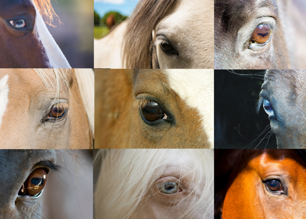
Horses have large, prominent eyes situated on either side of their head, as opposed to front-facing, like ours. This placement gives horses an impressive field of view, allowing horses to see an impressive 350 degrees around them! A grazing horse can see both the ground AND the horizon at the same time. Horses have both monocular vision, meaning they are able to see different things with each eye, and binocular vision, meaning they can focus on the same thing with both eyes. The placement of their eyes does have a disadvantage. They have blind spots directly behind and directly in front of them. (This is why it is important to approach horses carefully and ensure they can see you and they aren’t startled.)
Horses have, what we would consider, fairly good vision. Not 20/20 but still pretty good. Horses have lots (think millions!) of photoreceptors (light-sensitive cells) in their retinas. They can see colors, though in a more muted scheme than humans. Humans have 3 cones in their retinas while horses only have two. Horses are also able to see movement well, even in lower light. This is why a horse resident may “spook” at a sudden small movement from something seemingly unthreatening. This is a survival instinct that has served their feral and wild counterparts well. Their eyes, being large and prominent, are at risk of injuries, such as corneal ulcers, as well as other injuries and diseases. Now that we’ve taken a general look at vision in horses, let’s get into the anatomy.
General Anatomy Of The Eye
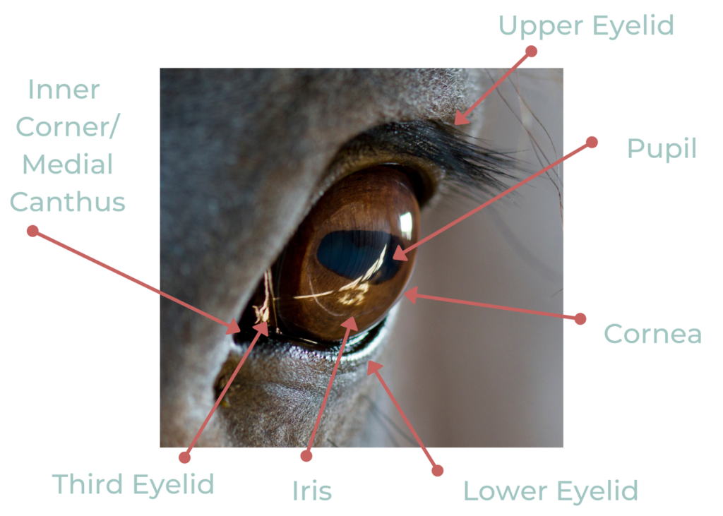
Let’s start out looking at the following basic diagram. This diagram covers superficial structures that you can readily see and identify. Being able to identify these structures and their roles will allow you to better understand your resident’s vision, provide better care, and communicate more effectively with an equine veterinarian.
Front-Facing View Of Eye
Glossary Of Terms
Cornea
The cornea is the clear, outer surface of the eye. It is a clear, dome-shaped structure that protects the eye and lets in light. It assists in focusing light on the retina, which sits at the back of the eye. The sclera, the white of the eye, lay beneath the cornea, as do the iris and pupil.
Inner Corner/Medial Canthus
The medial canthus is simply the inner corner of the eye.
Iris
The iris is the colored part of the eye. The iris is usually a shade of brown in horses, ranging from a lighter amber color to a deep, dark brown. However, some horses have blue irises, or, extremely rarely, green! The iris is also part of the uveal tract, which supplies the eye with blood.
Lower Eyelid
The lower eyelid is the structure of the skin at the bottom of the eye that helps spreads tears over the eye and keeps a protective moisture barrier and protects the eye from foreign objects.
Pupil
The pupil is the black horizontal oval area within the iris that enlarges or shrinks, in order to let in more or less light. If it is low light or night out, the pupil will enlarge to let more light in and if it is bright, the pupil will shrink to limit the amount of light.
Third Eyelid/Nictating Membrane
The third eyelid, or nictating membrane, is a thin mucous membrane that is pulled across the eye when a horse blinks. It is located at the inner corner of the eye. The third eyelid may pull up if the eye is inflamed or injured.
Upper Eyelid
The upper eyelid is the structure of skin above the eye that helps spreads tears over the eye and keeps a protective moisture barrier and protects the eye from foreign objects.
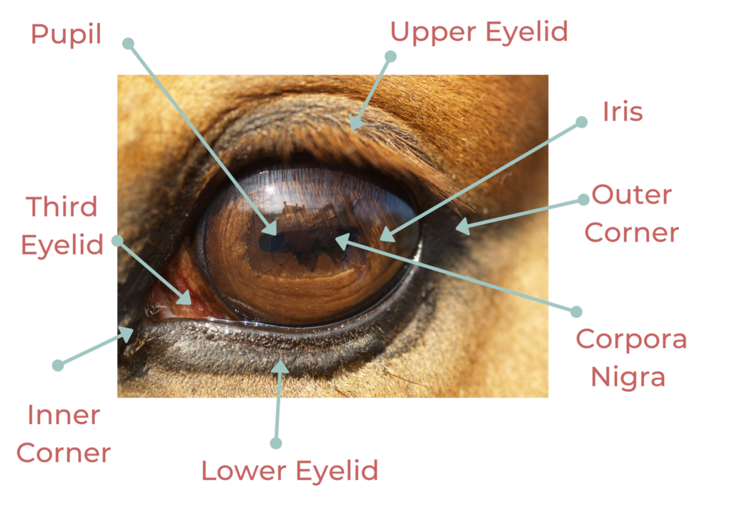
Side-Facing View Of Eye
Glossary Of Terms
Cornea
The cornea is the clear, outer surface of the eye. It is a clear, dome-shaped structure that protects the eye and lets in light. It assists in focusing light on the retina, which sits at the back of the eye. The sclera, the white of the eye, lay beneath the cornea, as do the iris and pupil.
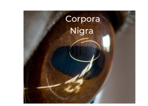
Corpora Nigra
The corpora nigra is the irregular-looking area of the iris dipping into the pupil. It may look a little like an inconsistent wavy pattern. It can also be seen at the bottom of the pupil in some horses. This structure is thought to help with glare from the light to help horses see better.
Inner Corner/Medial Canthus
The medial canthus is the inner corner of the eye.
Iris
The iris is the colored part of the eye. The iris is usually a shade of brown in horses, ranging from a lighter amber color to a deep, dark brown. However, some horses have blue irises, or, extremely rarely, green! The iris is also part of the uveal tract, which supplies the eye with blood.
Lower Eyelid
The lower eyelid is the structure of the skin at the bottom of the eye that helps spreads tears over the eye and keeps a protective moisture barrier and protects the eye from foreign objects.
Outer Corner (Lateral Canthus)
The lateral canthus is the outer corner of the eye.
Pupil
The pupil is the black horizontal oval area within the iris that enlarges or shrinks, in order to let in more or less light. If it is low light or night out, the pupil will enlarge to let more light in and if it is bright, the pupil will shrink to limit the amount of light.
Third Eyelid/Nictating Membrane
The third eyelid, or nictating membrane, is a thin mucous membrane that is pulled across the eye when a horse blinks. It is located at the inner corner of the eye. The third eyelid may pull up if the eye is inflamed or injured
Upper Eyelid
The upper eyelid is the structure of skin above the eye that helps spreads tears over the eye and keeps a protective moisture barrier and protects the eye from foreign objects.
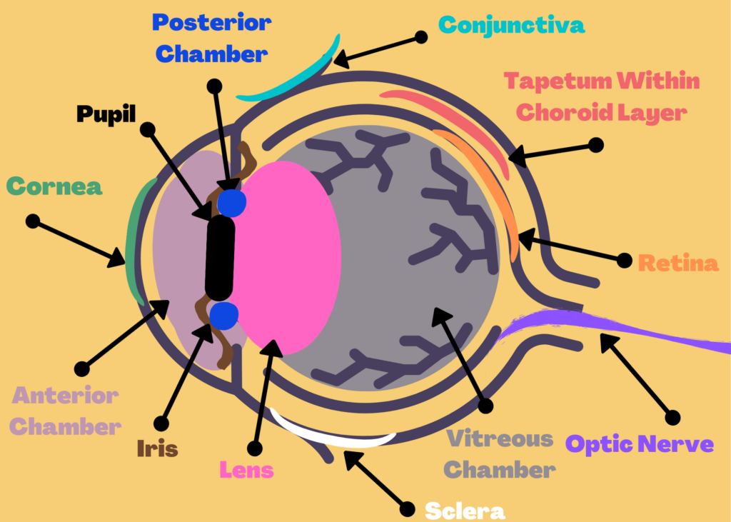
Inner Structures Of The Eye
Glossary Of Terms
Conjunctiva
The conjunctiva is a mucous membrane that covers the eye and lines the underside of the eyelid.
Anterior Chamber
The anterior chamber is the space between the cornea and the iris. It is fluid-filled.
Choroid
The membrane between the retina and the sclera (white of the eye). This membrane contains the tapetum, blood vessels, and pigmented cells.
Cornea
The cornea is the clear, outer surface of the eye. It is a clear, dome-shaped structure that protects the eye and lets in light. It assists in focusing light on the retina, which sits at the back of the eye. The sclera, the white of the eye, lay beneath the cornea, as do the iris and pupil.
Iris
The iris is the colored part of the eye. The iris is usually a shade of brown in horses, ranging from a lighter amber color to a deep, dark brown. However, some horses have blue irises, or, extremely rarely, green! The iris is also part of the uveal tract, which supplies the eye with blood.
Lens
The lens of the eye sits just behind the iris and changes its shape in order to focus light onto the retina. The lens may become thicker or thinner, depending on whether the horse is focusing on something close (it becomes thicker) or far away (it becomes thinner). The ciliary muscles are responsible for controlling the shape of the lens.
Optic Nerve
The optic nerve is the “channel” through which images are transmitted to the brain as electrical impulses.
Posterior Chamber
The posterior chamber is a small, fluid-filled chamber area behind the iris, next to the lens.
Pupil
The pupil is the black horizontal oval area within the iris that enlarges or shrinks, in order to let in more or less light. If it is low light or night out, the pupil will enlarge to let more light in and if it is bright, the pupil will shrink to limit the amount of light.
Retina
The retina is the inner layer of the back of the eye, which contains millions of light-sensitive cells, cones, and rods, that transmit what a horse is seeing through the optic nerve and into the brain.
Sclera
The sclera is the white of the eye.
Tapetum
The tapetum is reflective, located at the back of the eye, and helps improve vision in low light. This is the structure responsible for when we see “glowing” eyes from certain animals in the dark.
Vitreous Chamber
The vitreous chamber is the large area behind the lens, filled with a viscous (thick, sticky) substance.
Additional Glossary Terms
Cones
Cones are one of two types of photoreceptors (light-sensitive cells that turn images into electrical impulses to be sent to the brain via the optic nerve). Cones are necessary for sight in daylight and for distinguishing colors.
Rods
Rods are one of two types of photoreceptors (light-sensitive cells that turn images into electrical impulses to be sent to the brain via the optic nerve). Rods are light-sensitive cells that allow sight in dimmer light.
You made it! While this list is by no means an exhaustive one covering every structure in a horse’s eye, it should provide you with a foundational understanding. You can now use this knowledge to better understand and discuss eye health and potential eye health issues that might arise among horse residents. Of course, this resource is far from extensive and we recommend contacting your veterinarian regarding any specific eye health questions you may have. Stay tuned for an eye health and care resource!