




Malignant Catarrhal Fever (MCF) is a severe, often fatal systemic disease caused by a number of gammaherpesviruses belonging to the Macavirus genus. Each virus has a specific reservoir host species (sometimes referred to as the natural host or carrier host). While these reservoir species often harbor their respective virus, they typically do not develop clinical MCF. Susceptible non-reservoir hosts (sometimes referred to as incidental hosts), on the other hand, can develop serious, and even fatal, disease. Cows, American bison, water buffalo, deer, and other wild ruminant species are primarily affected by MCF, but it can also affect non-ruminant species, such as pigs.
Which Species Are Reservoir Hosts Of MCF-Causing Viruses?
The primary reservoir hosts of MCF-causing viruses are wildebeest, sheep, and goats, with almost all clinical cases being caused by wildebeest and sheep viruses, alcelaphine herpesvirus 1 (AlHV-1) and ovine herpesvirus 2 (OvHV-2), respectively.
AIHV-1 is endemic in wildebeest worldwide. It is believed that wildebeest calves become infected with AIHV-1 in utero or within the first few months of life. Infection of a non-reservoir host with AIHV-1 is often referred to as wildebeest-associated MCF or AIHV-1-associated MCF. Cases of AIHV-1-associated MCF have mostly been reported in sub-Saharan Africa, but they can also occur in other parts of the world where wildebeest and susceptible non-reservoir species are in close proximity (such as zoosOrganizations where animals, either rescued, bought, borrowed, or bred, are kept, typically for the benefit of human visitor interest. or farmsFor-profit organizations focused on the production and sale of plant and/or animal products. that house both wildebeest and susceptible species). European breeds of cows (Bos taurus, Bos indicus) are frequently affected, but cases have also been reported in banteng (Bos javanicus), bison (Bison sp), water buffalo (Bubalus bubalis), and African buffalo (Syncerus caffer).
Like AIHV-1 in wildebeest, OvHV-2 is endemic in sheep worldwide. Lambs typically become infected when they are between 3 and 9 months old. Outside of Africa, OvHV-2 is the major cause of MCF in domesticatedAdapted over time (as by selective breeding) from a wild or natural state to life in close association with and to the benefit of humans animals and accounts for the vast majority of cases in the US. While AIHV-1 is the most common cause of MCF in some African countries, disease from OvHV-2 has also been reported. Disease caused by OvHV-2 is often referred to as sheep-associated MCF or OvHV-2-associated MCF. American bison (Bison bison), banteng, and certain cervid species (including white-tailed deer and sika deer) are very susceptible to OvHV-2. Other susceptible species include European breeds of cows, water buffalo, pigs, goats, European bison (Bison bonasus), African buffalo, and giraffes.
As mentioned above, goats are also carriers of MCF viruses. Caprine herpesvirus-2 (CpHV-2) is endemic in most domesticated goat populations around the world, but compared to AIHV-1 and OvHV-2, far less is currently known about this virus. It has been reported to cause MCF in white-tailed deer, sika deer, moose, water buffalo, and certain antelope species. Goats can also be carriers of caprine herpesvirus-3 (CpHV-3, previously referred to as MCFV-WTD), which has been reported to cause MCF in deer in North America. There have also been reports of both asymptomatic and clinical OvHV-2 infections in goats, which we’ll discuss below.
Like other herpesviruses, MCF viruses cause lifelong, latent infections in reservoir hosts. While reservoir hosts do not typically develop clinical disease, rare cases of clinical OvHV-2 infection in sheep have been reported and are thought to result from unusually high virus replication.
How Do MCF Viruses Spread?
Infected reservoir hosts shed the virus primarily in their nasal and ocular secretions, but MCF-causing viruses have also been detected in feces and semen. Viral shedding is heaviest in younger individuals. Shedding of AIHV-1 peaks in wildebeest calves when they are 3-4 months old. Calves 6 months of age and older show lower levels of shedding, though the virus has been detected in the nasal secretions of older wildebeest who are stressed or who have been given corticosteroids (which are immunosuppressants). In lambs, viral shedding peaks between 6 and 9 months of age and decreases at 10 months. Intermittent shedding is possible in sheep of all ages, but adult sheep shed at a much lower rate than lambs. As mentioned above, MCF-causing viruses result in lifelong infection.
Currently, inhalation is believed to be the primary mode of transmission for MCF-causing viruses, but ingestion may also be possible. Susceptible non-reservoir hosts can become infected with MCF-causing viruses following direct or indirect contact with infected reservoir hosts. Pastures and fomitesObjects or materials that may become contaminated with an infectious agent and contribute to disease spread contaminated with MCF-causing viruses are believed to also play a role in transmission. While close contact between susceptible individuals and reservoir hosts is usually necessary for transmission, airborne transmission over long distances is possible. Transmission of AIHV-1 over a distance of 100 meters (109 yards) has been reported. In terms of airborne transmission of OvHV-2, there have been reports of transmission to cows separated from sheep by 70 meters (76.5 yards) and in bison separated by up to 5 km (3.1 miles) from a lamb feedlotA confined area where animals are kept and fed (typically high protein diets) in order for them to gain weight as quickly as possible.. However, in the latter case, it should be noted that a lamb feedlot would have the potential to be a place of significant viral shedding. Research suggests that in Canada, most outbreaks of OvHV-2 in bison occur when they are within 1 km (0.6 miles) of sheep.
Current evidence suggests that reservoir species are responsible for transmission both within their own species and to non-reservoir species. Non-reservoir species are primarily considered to be dead-end hosts (i.e., a sheep can transmit OvHV-2 to a cowWhile "cow" can be defined to refer exclusively to female cattle, at The Open Sanctuary Project we refer to domesticated cattle of all ages and sexes as "cows.", but that cow cannot transmit the virus to another cow they live with). However, both AIHV-1 and OvHV-2 can cross the placenta in cows, infecting calves in utero, and OvHV-2 has been detected in a water buffalo fetus. OvHV-2 nucleic acid has also been found in the secretions and excretions of some non-reservoir hosts, but, as of yet, it does not seem that there have been any definitive cases of horizontal spread from a non-reservoir species.
How Are Non-Reservoir Host Species Affected By MCF?
When a susceptible non-reservoir species is exposed to a reservoir host, they are at risk of MCF. According to The World Organization For Animal Health (WOAH), the species most often affected by MCF belong to the Cervidae (deer) family and Bovidae subfamily. However, as mentioned above, MCF has also been recognized in other species, including domesticated pigs, giraffes, and antelope species belonging to the Tragelaphinae subfamily. A particular non-reservoir species may be susceptible to one virus while showing some degree of resistance to others (i.e., a species may be highly susceptible to AIHV-1, but fairly resistant to OvHV-2 or vice versa). Additionally, different non-reservoir species show different degrees of susceptibility to these viruses.
Following exposure to an MCF-causing virus, the incubation period (the time between initial exposure and the onset of clinical signs) can vary depending on the specific virus, host, and other factors, but can range from 14 to over 200 days. However, not all susceptible non-reservoir hosts who are exposed to MCF-causing viruses will become sick. Seemingly healthy individuals can be seropositive (have detectable antibodies), and some studies have reported antibodies in up to 25-43% of healthy bison, cows, or pigs. There have also been reports of clinical cases of MCF developing in susceptible non-reservoir hosts who had been living with reservoir hosts for years without issue. Additionally, latent infection is possible in some species, and recurrence of clinical signs (recrudescence) following recovery is possible. All this could explain why there have been cases with no known exposure to reservoir species – the individual could have been carrying the virus for some time before showing clinical signs.
In general, it seems that younger non-reservoir hosts are affected more than older animals (but whether anything is known about truly elderly individuals is unclear since most information comes from non-sanctuary settings where animals typically do not have the opportunity to live out their natural lifespan). In some species, pregnant individuals may be more susceptible than other individuals.
Clinical cases of MCF can range from peracute to chronic, and in cases of subclinical disease, there is evidence to suggest that stress can bring about clinical illness. Below, we’ll look more closely at OvHV-2-associated MCF in key non-reservoir species.
Cows (Bos taurus and Bos indicus)
Cases of OvHV-2-associated MCF in cows are usually sporadic and affect a very small percentage of individuals in a herd (ranging from just one individual to a few). While morbidity is usually low, there have been rare outbreaks where up to 50% of a herd has been affected. Signs of clinical OvHV-2-associated MCF in cows are variable and can include nasal and ocular discharge, severe eye lesions, skin lesions (especially around the udders and perineum), enlarged lymph nodes, diarrhea (sometimes with blood in it), blood in urine, and/or lesions on the oral mucosa, which can result in excessive salivation due to oral discomfort. Neurological issues, such as incoordination and aggression, have also been reported. Bilateral corneal opacity that starts at the outer edge of the eye and progresses inward is common and is considered an important clinical sign suggestive of MCF. Cows with corneal opacity will appear sensitive to light and blind, which may be more obvious to caregivers than the changes to their eyes, especially if observing them from a distance. Affected individuals often also show other non-specific signs of illness, such as a high fever, inappetence, and depression.
The mortality rate in cows showing clinical signs of MCF averages between 80-90%. Compared to other species such as deer, American bison, and water buffalo, cows tend to have a more prolonged period of illness before succumbing to the disease. Individuals who recover may have lasting eye lesions. As mentioned above, recrudescence of clinical signs is possible in those who recover.
American Bison
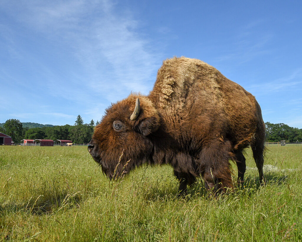
American bison are highly susceptible to OvHV-2. In fact, they are more than 1,000 times more susceptible to infection than cows. European bison are also susceptible to OvHV-2 infection, but less so than American bison. In American bison herds with close contact to sheep or where individuals are stressed, morbidity can reach 50-100%, resulting in large outbreaks. In 2003, an outbreak at a feedlot killed 825 American bison following exposure to sheep. Infection often results in peracute disease, with individuals dying without first showing clinical signs of disease. When signs are noted, they typically include respiratory, ocular, and gastrointestinal signs similar to those commonly seen in cows, and neurological signs are also possible. Individuals often hide signs of illness until they are close to death. Comparatively, the presence of blood in feces and urine is more common in bison than in cows, and lymph nodes are typically less enlarged in bison than in cows. In clinical cases, mortality can reach 100%.
Water Buffalo
Water buffalo are typically described as being more susceptible to OvHV-2 infection than cows but less susceptible than American bison. Clinical signs are similar to those seen in cows, but corneal opacity is less common in water buffalo. More often, they develop mild to moderate conjunctivitisConjunctivitis is the inflammation of the conjunctiva of the eye.. Mortality rates can be as high as 75-100% in clinical cases.
In addition to OvHV-2, there have also been reports of MCF in water buffalo caused by CpHV-2.
Pigs
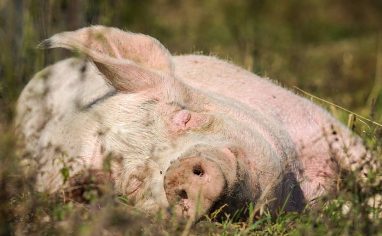
Pigs are also susceptible to OvHV-2, and sporadic cases, including a few outbreaks, have been reported. Compared to cases in ruminants, confirmed cases in pigs are currently rare, but we are aware of at least one case involving a sanctuary pig resident. Pigs typically develop acute or peracute disease, but chronic cases lasting several weeks have also been reported. In acute cases, individuals commonly develop fever and labored breathing. Other clinical signs reported in pigs include malodorous nasal discharge, nasal and oral lesions, eye issues, blood in urine, skin lesions, neurologic signs, fever, inappetence, and lethargy. In one outbreak, 41 of 120 pigs exposed to lambs developed clinical disease and, of the 41 affected individuals, 19 died.
Goats
As mentioned above, in addition to being reservoir hosts for CpHV-2 and CpHV-3, there have been reports of both asymptomatic and clinical cases of OvHV-2 in goats. Further research is needed to determine the prevalence of OvHV-2 in healthy goats, but a study conducted in Pakistan found OvHV-2 DNA in blood samples from 22 out of 50 healthy goats, and testing of 6 healthy goats in Switzerland detected OvHV-2 in samples from just one goat. While their ability to transmit CpHV-2 and CpHV-3 to susceptible non-reservoir species (e.g., white-tailed deer, sika deer, moose, water buffalo, and certain antelope species) has been confirmed, whether or not goats can also transmit OvHV-2 to others seems to be unclear as of the time of this writing.
For the most part, clinical cases of OvHV-2-associated MCF in goats seem to be rare, especially compared to some of the other susceptible species, but they have been reported. Clinical signs have included fever, nasal discharge, diarrhea, chronic weight loss, corneal opacity, alopeciaAlopecia is the loss of hair, wool, or feathers., skin lesions, and neurological signs.
Other Species Of Note
Other species have shown some susceptibility to MCF-causing viruses. While we are fervently against animal experimentation, it’s important to note that rabbitsUnless explicitly mentioned, we are referring to domesticated rabbit breeds, not wild rabbits, who may have unique needs not covered by this resource., cavies (guinea pigs), rats, and Syrian hamsters have been experimentally infected. Additionally, a possible clinical case of OvHV-2-associated MCF was reported in an alpaca, and OvHV-2 nucleic acids were found in a sick foal and an asymptomatic horse.
What Diagnostic Tests Are Available?
Several diagnostic tests are available to detect MCF-causing viruses. PCR testing detects viral DNA and is recommended to confirm an MCF diagnosis in a clinical non-reservoir species. As mentioned earlier, clinical cases of MCF in reservoir species are rare but not unheard of. Unfortunately, antemortem confirmation of MCF in a clinical reservoir host is difficult. Because infection is endemic in reservoir host species, detecting viral DNA or antibodies is not diagnostically significant and cannot definitely confirm that the virus is the cause of clinical signs. However, the presence of high levels of viral DNA in tissues can be used to support a suspected diagnosis.
Antibody testing (e.g., ELISA) can be performed in reservoir hosts to identify carriers. However, maternal antibodies can interfere with testing in lambs who are younger than 1 month old, so your veterinarian may recommend waiting to test young lambs. It can take more than 4 weeks for individuals to produce detectable levels of antibodies after being exposed to a low dose of virus, so repeat testing may be recommended following a negative result.
Antibody testing may also be used to identify subclinical infections in susceptible non-reservoir species, but antibody testing cannot be used to confirm or rule out MCF in a clinical individual. One reason for this pertains to the amount of time it may take for the individual to produce detectable levels of antibodies. An individual could become very ill and even die from MCF before producing antibodies that would lead to a positive test result. Therefore, a negative antibody test cannot rule out MCF. Additionally, because antibodies have been detected in healthy susceptible species (indicating latent or subclinical infection), the presence of antibodies alone cannot confirm that an MCF-causing virus is the cause of an individual’s clinical signs. Instead, PCR should be performed in clinical patients because clinical cases of MCF result in high levels of viral DNA, whereas latent and subclinical infections result in little to no viral DNA. If a resident passes away or is euthanized and MCF is suspected, a post-mortem examination can be performed to confirm the diagnosis.
In the US, MCF is a monitored disease in bovines. Monitored diseases are endemic diseases that are tracked by the Animal and Plant Health Inspection Service (APHIS) using data provided by State Animal Health Officials and labs. In addition to federal regulations, reportable diseases can vary by state. Whether you are in the US or elsewhere, your veterinarian will be able to tell you if MCF is reportable in your region.
Are There Any Treatment Options For MCF?
Unfortunately, there is currently no treatment for MCF. All veterinarians can do is attempt to manage symptoms and provide supportive care.
What Can Sanctuaries Do To Prevent MCF?
With no treatment options or commercial vaccines currently available, prevention is key. The only effective way to reduce the risk of exposure to MCF-causing viruses is to maintain separation between susceptible non-reservoir hosts and potential carriers. The distance needed to prevent airborne transmission is currently unknown and likely affected by numerous factors such as the degree of susceptibility of the non-reservoir host, the amount of virus being shed by reservoir hosts, the number of individuals shedding, and environmental conditions. Given the reports of transmission occurring over significant distances, folks should provide as much separation as possible between susceptible non-reservoir and reservoir hosts.
Generally speaking, the following practices can help reduce the risk of MCF in susceptible non-reservoir species.
- If you care for both reservoir species and susceptible non-reservoir species, house them separately.
- Keep as much distance as possible between susceptible non-reservoir hosts and reservoir hosts, focusing on providing the most distance and taking extra biosecurity measures to protect your most susceptible residents.
- Consider exposure risks outside the sanctuary (such as a neighboring sheep farm), and take steps to increase distance from those risks as well.
- Do not let susceptible non-reservoir species graze on pastures recently grazed by reservoir species.
- Enact biosecurityMerck Veterinary Manual defines biosecurity as ”the implementation of measures that reduce the risk of the introduction and spread of disease agents [pathogens].” measures that reduce the risk of transmission via fomites or mechanical transmission. We recommend working with your veterinarian to determine which specific measures are most appropriate for your specific situation.
- Remember that lambs (and wildebeest calves) are the biggest shedders of MCF-causing viruses. Depending on the species you care for, your philosophy of care, and the size of your sanctuary, you may determine that it is not safe to rescue lambs and house them on sanctuary grounds. If you decide to take in lambs, be very mindful of where they are housed in relation to susceptible individuals, providing as much distance and separation as possible, and enact biosecurity measures to reduce the risk of transmission via fomites.
- Caregivers should always take steps to keep stress levels at a minimum for residents, but this can also help with MCF prevention since stress has been shown to play a role in subclinical cases becoming clinical and could also play a role in viral shedding of reservoir hosts.
If you care for susceptible non-reservoir species, we strongly encourage you to talk to your veterinarian about specific ways you can reduce your residents’ risk of MCF. This is always a good idea, but it is critical for diseases like MCF that still have so many unknowns. Because OvHV-2 is endemic in sheep, testing to identify carriers may not be recommended. Instead, your veterinarian may recommend acting as if all your sheep residents are potential carriers. If you care for multiple susceptible non-reservoir species, your veterinarian may recommend different biosecurity measures for each species based on their degree of susceptibility (for example, given how highly susceptible bison are to OvHV-2, your vet may recommend more stringent biosecurity measures for them than for species that are more resistant). This is not to say that your other susceptible residents do not require biosecurity measures to reduce the risk of exposure, but the steps you take to protect each species may look different due to differences in their degree of susceptibility.
MCF is a challenging and unpredictable disease. Given that farmed animalA species or specific breed of animal that is raised by humans for the use of their bodies or what comes from their bodies. sanctuaries often care for both reservoir and susceptible non-reservoir species, it’s important that they are aware of this disease. By understanding how it is transmitted and working with your veterinarian to establish appropriate biosecurity measures, you can take important steps towards reducing your residents’ risk as much as possible.
SOURCES:
Malignant Catarrhal Fever In Animals | Merck Veterinary Manual (Non-Compassionate Source)
Malignant Catarrhal Fever | World Organization For Animal Health (Non-Compassionate Source)
Malignant Catarrhal Fever | Center For Food Security And Public Health (Non-Compassionate Source)
Diagnosis And Testing For Malignant Catarrhal Fever (MCF) | Washington Animal Disease Diagnostic Laboratory (Non-Compassionate Source)
Two Different Macaviruses, Ovine Herpesvirus-2 And Caprine Herpesvirus-2, Behave Differently In Water Buffaloes Than In Cattle Or In Their Respective Reservoir Species | Stahel AB, Baggenstos R, Engels M, Friess M, Ackermann M. (Non-Compassionate Source)
A Devastating Outbreak Of Malignant Catarrhal Fever In A Bison Feedlot | Li H, Taus NS, Jones C, Murphy B, Evermann JF, Crawford TB. (Non-Compassionate Source)
Hide And Seek | US Department Of Agriculture (Non-Compassionate Source)
An Outbreak Of Porcine Malignant Catarrhal Fever In A Farrow-To-Finish Swine Farm In The United States | Gauger PC, Patterson AR, Kim WI, et al. (Non-Compassionate Source)
Granulomatous Mural Folliculitis And Cytotoxic Interface DermatitisDermatitis refers to skin irritation. This may include itchy, dry skin or a rash on swollen, reddened skin. It may also cause the skin to blister, ooze, crust or flake In A Pygmy Goat Associated With Ovine Herpesvirus-2 And Systemic Lesions Of Malignant Catarrhal Fever | Veterinary Case Reports (Non-Compassionate Source)
Molecular Detection And Characterization Of Ovine Herpesvirus-2 Using Heminested PCR In Pakistan | Riaz A, Dry I, Dalziel R, Rehman SU, Shah MA, Akhtar HMN, Yousaf A, Baig R. (Non-Compassionate Source)